The Pyloric Sphincteric Cylinder in Health and Disease


Go to chapter: 1 | 2 | 3 | 4 | 5 | 6 | 7 | 8 | 9 | 10 | 11 | 12 | 13 | 14 | 15 | 16 | 17 | 18 | 19 | 20 | 21 | 22 | 23 | 24 | 25 | 26 | 27 | 28 | 29 | 30 | 31 | 32 | 33 | 34 | 35 | 36 | 37 | 38 | 39
Chapter 33 (page 168)


Results

Duodenum

Radiographic Appearance of Duodenal Bulb Radiographically the base of the duodenal bulb, as well as its other walls and lumen appeared normal in 40 of 50 cases of pyloric carcinoma, showing no evidence of tumor extension (Fig. 33.1). In 2 of the remaining 10 cases a concave, smooth indentation of the base of the bulb, without other signs of duodenal involvement, was seen (Fig. 33.5). In 2 cases smaller, concave indentations of the base of the bulb on either side of the pyloric aperture were noted (Fig. 33.3). Between these indentations a small, triangular projection of barium extended into the aboral side of the pyloric aperture; this we have called the duodenal "tail" (Chap. 13). There was a suggestion of infiltration of the base of the duodenal bulb in 2 of the 10 cases. In the remaining 4 cases the bulb was not visualized adequately owing to technical factors (e.g. overlying partially obstructed stomach) and possible duodenal extension could not be determined.

Incidence of Duodenal Spread as Determined at Operation An attempt was made to determine duodenal involvement or non-involvement by inspection and palpation in 44 cases at operation. In 9 it was not possible to evaluate the pyloro-duodenal junction adequately on account of the tumor mass itself, infiltration of surrounding structures, liver metastases and/or ascites (Table 33.2); these cases were exluded. In other words, 35 cases could be evaluated.

Table 33.2

Type and number of operationsCases without macroscopic duodenal involvement Cases with macroscopic involvementUnevaluated

B II242031
B I2200
Gastroenterostomy16628
Laparotomy2200

TOTAL443059


In 23 evaluated Billroth II partial gastrectomies there was no evidence of macroscopic duodenal spread in 20; duodenal involvement was thought to be present in 3. In 2 Billroth I partial gastrectomies and 2 exploratory laparotomies no duodenal extension was evident. In 8 evaluated palliative gastroenterostomies the duodenum appeared to be uninvolved in 6 cases and duodenal extension was noted in 2. In other words, macroscopic duodenal spread was thought to be present in 5 of 35 cases evaluated at operation. (Histology subsequently disproved duodenal spread in one of the 5 cases; in the 2 cases with gastroenterostomies spread was not confirmed owing to lack of histological examination. In 3 of the 30 cases in which the duodenum appeared normal at operation, histology did show duodenal spread). It is confirmed that at operation the duodenum may appear normal macroscopically, in the presence of microscopic evidence of transpyloric spread of pyloric adenocarcinoma.

Incidence and Extent of Duodenal Spread as seen Microscopically Histological examination was obtained in 23 resection specimens. Of these 16 showed no evidence of duodenal extension. In 7 cases duodenal spread of pyloric carcinoma was seen microscopically; these will be described briefly.

Case Reports

Case 33.8 J.P., 39 year old male. Radiology: 5.0cm long constricting pyloric filling defect. Smooth, concave indentation base of duodenal bulb (Fig. 33.8). Operation: Entire lesser curvature of stomach, part of greater curvature and pyloric region infiltrated. Serosal spread, lymph node and probable liver metastases. Duodenum appears normal. Billroth II. Gastric histology: well differentiated adenocarcinoma with widespread infiltration of gastric submucosa and mucosa. Duodenal histology: spread into submucosa and to lesser extent into mucosa of proximal 2.0 cm of duodenum.

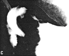
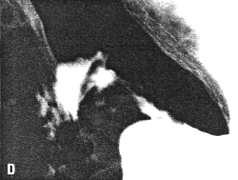
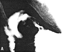
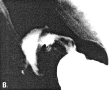
Fig. 33.8 A-D. Case J.P. Constricting pyloric filling defect. Smooth, concave indentation base of duodenal bulb.


Previous Page | Table of Contents | Next Page
© Copyright PLiG 1998