

Go to chapter: 1 | 2 | 3 | 4 | 5 | 6 | 7 | 8 | 9 | 10 | 11 | 12 | 13 | 14 | 15 | 16 | 17 | 18 | 19 | 20 | 21 | 22 | 23 | 24 | 25 | 26 | 27 | 28 | 29 | 30 | 31 | 32 | 33 | 34 | 35 | 36 | 37 | 38 | 39
Chapter 32 (page 155)
Chapter 32
Gastro-oesophageal Reflux Disease (GERD) and the Pyloric Sphincteric Cylinder
Roviralta (l951) described 3 cases of partial thoracic stomach in infants associated with
hypertrophic pyloric stenosis (IHPS), and called the combination the phreno-pyloric
syndrome. It was believed that raised intragastric pressure secondary to the obstruction at
the pylorus forced the stomach into the chest. Among 115 children with a partial thoracic
stomach, Astley and Carré (l954) encountered 5 who also had hypertrophic pyloric
stenosis, while another 3 had "infantile pylorospasm". The pylorospasm in all 3 cases
was described as an inconstant narrowing of the pyloric "antrum", (Chap. 20), the
radiographic appearance simulating IHPS to such a degree that Astley (l956) later called
them cases of pseudo-hypertrophic pyloric stenosis. The symptomatology suggested that
initially there was gastroesophageal reflux due to a partial thoracic stomach, followed by
the superimposition of hypertrophic stenosis a few weeks later. Thus vomiting
commenced soon after birth, and at the age of 2 to 3 weeks the symptoms and signs of
hypertrophic stenosis, such as projectile vomiting, visible peristalsis and a palpable mass
were superadded.
Forshall (l955) described the findings in 93 infants with gastroesophageal reflux and
hiatus hernia. In 58 cases the cardia was incompetent but situated below the diaphragm.
Eight of these required Ramstedt's operation for IHPS, while others had visible gastric
peristalsis with temporary palpable masses in the pyloric region.
Astley (l956) stated that the association of hiatus hernia and hypertrophic pyloric stenosis
was not a very common combination, but that the frequency was enough to suggest
something more than a chance occurrence. He found no ready explanation for the
association of these two conditions.
Stewart (l960), in discussing a paper by Herrington (l960), was impressed by the
frequency of pyloric hypertrophy in cases of hiatus hernia; in many instances it
resembled infantile hypertrophic pyloric stenosis.
Johnston (l960), in a series of 76 cases of hiatus hernia in childhood, found that 8 (10.5
percent) also had hypertrophic pyloric stenosis. Some of those without hypertrophic
stenosis showed visible gastric peristalsis with forcible or even projectile vomiting, which
to him was an indication of a gastric emptying disorder, giving rise to functional pyloric
obstruction. It was reasoned that this raised the intragastric pressure, thus forcing the
cardia into the chest.
Bowen (l988) pointed out that criteria for diagnosing hiatus hernia in infants remained
unsettled, but that it was generally agreed that the retrograde passage of material from the
stomach into the oesophagus was the crux of the matter, regardless of whether or not a
hiatal hernia could be demonstrated convincingly.
In a number of infants we have noted a combination of hiatus hernia and IHPS; however,
no systematic study was done in infants to determine in which percentage of hiatus hernia cases
IHPS also occurred. The following is an example of one of our cases:
Case Reports
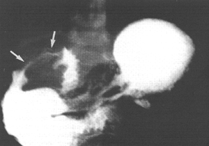
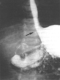
Case 32.1. E.B., 5 weeks old female infant, was admitted with a history of vomiting
after feeds and recurrent bilateral pneumonia. Radiographic examination showed a
severe, constant narrowing of the pyloric sphincteric cylinder, with a "string sign" typical
of IHPS (Fig. 32.1A). The gastro-oesophageal junction was patulous with
free and persistent gastro-oesophageal reflux, diagnosed radiographically as a sliding
hiatus hernia (Fig. 32.1B). Some aspiration of refluxed barium occurred. At
operation the next day a pyloric "olive" measuring approximately 2.3 cm x 0.8 cm,
typical of IHPS, was found. Ramstedt pyloromyotomy was done; post-operatively
vomiting stopped and the patient made an uneventful recovery.
 A A | |
| Fig. 32.1 A,B.
Case E.B. A Constant narrowing of pyloric sphincteric cylinder with string sign (arrows),
typical of idiopathic hypertrophic pyloric stenosis.
B Patulous gastro-oesophageal junction with free reflux (arrow)
|  B B |
Previous Page | Table of Contents | Next Page
© Copyright PLiG 1998


 A
A B
B