The Pyloric Sphincteric Cylinder in Health and Disease


Go to chapter: 1 | 2 | 3 | 4 | 5 | 6 | 7 | 8 | 9 | 10 | 11 | 12 | 13 | 14 | 15 | 16 | 17 | 18 | 19 | 20 | 21 | 22 | 23 | 24 | 25 | 26 | 27 | 28 | 29 | 30 | 31 | 32 | 33 | 34 | 35 | 36 | 37 | 38 | 39
Chapter 23 (page 103)


Chapter 23

Infantile Hypertrophic Pyloric Stenosis

Although a few isolated cases of infantile hypertrophic pyloric stenosis (IHPS) had been reported previously, Hirschsprung (l888) is generally regarded as the first author to have recognized it as a separate clinical entity. At that time the tendency was to designate the condition congenital hypertrophic pyloric stenosis. At present, the clinical features and management are well understood, and will not be recapitulated. It may be useful to discuss the images seen during radiographic and ultrasonic examinations, especially in relation to anatomical factors, and to consider some of the theories regarding the etiology and pathogenesis.

Anatomical Localization and Radiographic Features

Cunningham (1906) studied the morbid anatomical appearances in "a considerable number" of cases of IHPS, and found the muscular hypertrophy to be limited to the anatomical pyloric sphincteric cylinder, which includes the pyloric ring (Chap. 3). The affected area was almost 2.54 cm in length and had the look and feel of a hard, solid cylinder. It was sharply demarcated from the duodenum on its aboral, and from the pyloric vestibule on its oral side. In more severe cases the narrowing was greatest at the two ends, the cylinder in those cases assuming an oval or fusiform shape, somewhat like an olive. In all cases the musculature of the remainder of the stomach was normal. The relative extent to which each of the two muscular layers was involved in the hypertrophy was not the same in every case. While some observers believed that the circular musculature had undergone the greater degree of hypertrophy, Cunningham (1906) found that both layers were involved. Nevertheless the circular layer of the cylinder remained 3 to 4 times as thick as the longitudinal in his cases.

Forssell (l913) emphasized that muscular hypertrophy in cases of IHPS was localized to the canalis egestorius, i.e. the pyloric sphincteric cylinder of Cunningham (1906) (Chap. 3). The radiographic appearances of the contracted canalis in IHPS corresponded exactly to those of a maximal normal contraction of the pyloric sphincteric cylinder (Chap. 13); while the contraction was cyclical and of a fleeting nature in normal subjects, it was constant in IHPS. The muscular hypertrophy was associated with functional abnormalities of the closing mechanism of the canalis egestorius. The autoplastic movements of the mucous membrane were also involved (Chaps. 2, 13).

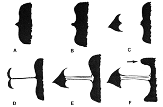
Meuwissen and Sloof (l932, l934) were the first to state that the purpose of the radiological examination was not to demonstrate the indirect features of the condition, such as gastric hyperperistalsis and retention, but to visualize the lesion itself, i.e. the contracted pyloric canal, in its longest dimension. The average length of the canal in normal infants was only 1.0 to 2.0 mm, while in the majority of cases of IHPS it was very long and narrow, ranging from 12.0 to 24.0 mm in length. In a minority of cases the range was from 3.0 to 11.0 mm. (Owing to magnification factors the actual length was about nine-tenths of that measured on the films). Radiologically the thin, permanently contracted canal, containing a single, central streak of barium in the lumen, resembled a string. These authors were the first to describe the "string sign" of IHPS, and also mentioned concave indentations of the distal stomach on either side of the string, caused by the bulging pyloric musculature, which became known as the "shoulder sign" (Fig. 23.1). There was complete absence of peristalsis in the contracted region. According to Meuwissen and Sloof (l932, l934) the contraction was largely due to spasm, with or without associated muscular hypertrophy.

Fig. 23.1 A-F. More common radiographic signs of infantile hypertrophic pyloric stenosis (after Astley, 1952). A Central "beak". B Beak with adjacent concave indentations (shoulder sign). C Beak, gap and cap. D String sign. E Longitudinal mucosal folds. F Concave indentation base of cap. Pyloric "tit" (arrow)


Previous Page | Table of Contents | Next Page
© Copyright PLiG 1998