


Go to chapter: 1 | 2 | 3 | 4 | 5 | 6 | 7 | 8 | 9 | 10 | 11 | 12 | 13 | 14 | 15 | 16 | 17 | 18 | 19 | 20 | 21 | 22 | 23 | 24 | 25 | 26 | 27 | 28 | 29 | 30 | 31 | 32 | 33 | 34 | 35 | 36 | 37 | 38 | 39
Chapter 25 (page 118)
Chapter 25
Focal Hypertrophy and Focal Spasm of the Pyloric Musculature in Adults
In l952 Bachmann reported 12 cases with localized areas of hypertrophy of the pyloric
musculature in adults. In each instance the hypertrophied area was situated on the lesser
curvature; it was found as incidental pathology in a series of 600 autopsies and none of
the cases had any other abnormalities of the stomach and duodenum. The cases were
divided into three groups according to their relationship to the pyloric "sphincter".
(Comment: the "sphincter" was equated with the pyloric ring). In the first
group (5 cases) the thickening was situated directly in the "sphincter"; it resembled an
enlarged version of the normal sphincter, consisting mainly of circular but also
containing some irregular and longitudinal muscle fibres. In the second group (4 cases)
the thickening was located a very short distance orally to the "sphincter", being separated
from it by a narrow zone of normal tissues. In the third group (3 cases) it was situated in
the "sphincter" as well as in the immediate prepyloric part. In the first two groups it
appeared to be of a rounded or nodular character (while differing distinctly from a
myoma), and in the third group it was rather longer than wide. In considering the
pathogenesis, Bachmann asked himself whether these cases indicated that a certain part
of the pyloric musculature was liable to undergo hypertrophy by virtue of its having a
specialized function or structure.
Keet (l956) reported 2 adult operative cases in which the gastric resection specimens
showed, as incidental pathology, a localized area of thickening of the pyloric musculature
on the lesser curvature of the stomach. In the first case a true muscular hypertrophy was
present; in the second the thickening appeared to be in the nature of a spasm, as it
disappeared during the course of a few hours. We believe that the localized or focal
pyloric muscular thickening in these two cases lends itself to a rational explanation on
anatomical grounds, as indicated below.
Case 25.1. G.V.L., 50 year old male, was admitted for partial gastrectomy because of
a non-healing gastric ulcer, having had ulcer symptoms for the previous 12 years.
Radiographic examination 6 years prior to admission had shown a tiny excrescence on
the lesser curvature of the stomach 1.5 cm proximal to the pylorus, which was interpreted
as a gastric ulcer. Three years prior to admission a second radiographic examination
reported an ulcer niche on the lesser curvature, but failed to state its exact situation. Four
months before admission a third radiographic examination showed a large gastric ulcer
niche halfway up the lesser curvature in the region of the incisura angularis; there was no
sign of the ulcer previously mentioned proximal to the pylorus. At operation the gall
bladder was distended and contained calculi; there were no adhesions to the duodenum
or pylorus. Palpation of the stomach in situ revealed a thickening of softish consistency
in the pylorus on the lesser curvature side, diagnosed provisionally as a gastric polyp. A
large gastric ulcer of the middle of the lesser curvature was seen and felt. Partial
gastrectomy was done, the duodenum being divided 3.0 cm distal to the pyloric ring, well
beyond the palpable thickening. A retrocolic gastrojejunal anastomosis was made and a
cholecystectomy performed.
The macroscopic pathological examination of the resection specimen showed a large
gastric ulcer on the lesser curvature 7.0 cm proximal to the pylorus. A mucosal fold,
approximately l.0 cm high, separated the lumen of the stomach from that of the
duodenum. In the pylorus, on the lesser curvature side and jutting into this mucosal fold,
a rounded, pea-sized, rubbery hard mass was situated in the gastric wall. The mucosa,
which was freely mobile on the underlying layers, was less mobile over the mass. There
was no naked-eye evidence of ulceration locally. Microscopic examination showed the
mass to consist of hyperplasia of the circular muscle (Fig. 25.1); it was not a myoma as it
was not well defined and merged gradually into the surrounding circular muscle. The
submucosa overlying the thickening was rather thin and contained numerous blood
vessels. The pyloric mucosa and submucosa showed infiltration with inflammatory cells
and changes of chronic gastritis. The ulcer on the middle of the lesser curvature proved
to be a chronic, benign ulcer penetrating into the muscle layers.
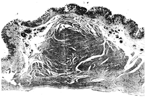 |
Fig. 25.1.
Case G.V.L. Microscopic section of pyloric nodule on lesser curvature side of
pyloroduodenal junction, showing great hypertrophy of circular muscle fibres. Thin
overlying submucosa
|
Case 25.2 M.B., 58 year old female, was admitted because of achylia gastrica.
Radiographic examination showed an irregularity on the lesser curvature at the incusura
angularis, which was regarded as a probable early gastric carcinoma. Partial gastrectomy
was performed. The macroscopic examination of the fresh resection specimen showed a
few hemorragic spots in its proximal part. At the pylorus, on the lesser curvature side, a
hard pea-sized nodule was felt in the gastric wall. It was not particualrly well defined
and was presumed to be a local thickening of the pyloric musculature. Microscopic
examination showed well marked chronic gastritis with a few erosive defects in the
mucosa and fibrotic tissue in the submucosa. When the specimen was handled again a
few hours after it had been received, it was noted that the nodule previously felt at the
pylorus on the lesser curvature side had disappeared. Except for the chronic gastritis no
microscopical abnormalities were seen in the pylorus.
Previous Page | Table of Contents | Next Page
© Copyright PLiG 1998