The Pyloric Sphincteric Cylinder in Health and Disease


Go to chapter: 1 | 2 | 3 | 4 | 5 | 6 | 7 | 8 | 9 | 10 | 11 | 12 | 13 | 14 | 15 | 16 | 17 | 18 | 19 | 20 | 21 | 22 | 23 | 24 | 25 | 26 | 27 | 28 | 29 | 30 | 31 | 32 | 33 | 34 | 35 | 36 | 37 | 38 | 39
Chapter 8 (page 29)


Chapter 8

Nerves

Classically the autonomic nervous system consists of two components, cholinergic and adrenergic nerves; the former are contained mostly in the parasympathetic, and the latter mostly in the sympathetic division. For the past 4 decades a third component of the autonomic system, which is neither cholinergic nor adrenergic, has been recogized; the principal active substance released from these nerves in the gastrointestinal tract is a purine nucleotide; they have been called purigenic nerves by Burnstock (l972). An increasing number of peptides with a dual localization in endocrine cells and peripheral nerves in the walls of the gastrointestinal tract has now been recognized. This has led to the concept of a three-part autonomic control system consisting of cholinergic, adrenergic and peptidergic nerves (Bishop et al. l977).

Parasympathetic Nerve Supply

The parasympathetic nerve supply to the stomach is derived from the anterior and posterior vagal trunks and their branches. Afferent fibres are also present in the vagi, which are mixed nerves.

Anterior Vagus

The anterior vagal trunk, which is derived mainly from the left vagus nerve but which includes fibres from the right vagus and also some sympathetic fibres from the splanchnic nerves, enters the abdominal cavity through the oesophageal hiatus in the diaphragm. It is usually single but may be divided into two or even three trunks. The manner of distribution of its branches is in the main constant (Latarjet et al. l921; McCrea l924).

Having supplied several fine twigs to the lower end of the oesophagus and cardiac part of the stomach, the anterior trunk breaks up into its main branches.

According to Latarjet three main sets of branches are present:
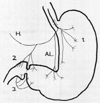
  1. The first set, consisting of 4 to 5 direct branches, emanating "one below the other", supplies the upper part of the lesser curvature. These nerves do not form a plexus and can be dissected separately. A few filaments from the sympathetic supply join these direct branches via the coeliac plexus. One of the branches in this group is always bigger than the others and very distinct; Latarjet (l921) called it the principal anterior nerve of the lesser curvature. It innervates the area from the cardia to the pyloric vestibule, and does not proceed beyond the latter (Fig 8.1).

  2. Branches emanating from the vagal supply to the liver. These branches, usually 3 to 5 in number, descending in the lesser omentum, "fall like rain" on the superior margin of the pylorus and first part of the duodenum. Latarjet called them the superior pyloric nerves (Fig 8.1).

  3. The third set consists of vagal filaments from the hepatic branches, accompanying the sympathetic nerves along the right gastroepiploic artery next to the greater curvature. They provide a few vagal twigs to the inferior margin of the pylorus. Latarjet called these the inferior pyloric nerves (Fig 8.1).


According to Latarjet (l921) the anterior vagus can be divided into two distinct functional divisions. The first division, consisting of the direct branches, supplies the fornix and body, i.e. the "reservoir" part of the stomach. The second division, through the hepatic branches, supplies the pylorus and first part of the duodenum, i.e. the "sphincteric" part of the stomach. (Comment: According to Latarjet, the principal anterior nerve of the lesser curvature does not supply the pylorus; the pylorus and first part of the duodenum are innervated by the superior and inferior pyloric nerves, emanating from the hepatic branches). Latarjet found the innervation in dogs to be similar to that in man.

The papers of McCrea (l924), Mitchell (l940) and Jackson (l948) describe personal investigations and provide an analysis of the literature. The following findings are pertinent to the present discussion.

According to McCrea (l924) the anterior vagal trunk breaks up into 3 to 6 main branches. To the left a group of gastric branches arise, supplying the anterior surface of the cardia, the fornix and proximal portion of the body of the stomach. These may or may not form an anterior gastric plexus.

Fig. 8.1. Diagram of gastric branches of anterior vagus. 1., Direct branches; 2., branches emanating from vagal supply to liver (superior pyloric nerves); 3., branches emanating from vagal supply to liver (inferior pyloric nerves); H., hepatic branch or branches; A.L., anterior nerve of Latarjet (principal anterior nerve of lesser curvature)


Previous Page | Table of Contents | Next Page
© Copyright PLiG 1998