


Go to chapter: 1 | 2 | 3 | 4 | 5 | 6 | 7 | 8 | 9 | 10 | 11 | 12 | 13 | 14 | 15 | 16 | 17 | 18 | 19 | 20 | 21 | 22 | 23 | 24 | 25 | 26 | 27 | 28 | 29 | 30 | 31 | 32 | 33 | 34 | 35 | 36 | 37 | 38 | 39
Chapter 26 (page 121)
Chapter 26
Nausea, Retching and Vomiting
The mechanism of vomiting in mammals is complex and in spite of experimental studies
some aspects are still not fully understood. It is usually accepted that the vomiting
sequence consists of 3 successive phases: nausea initially, followed by retching, often
leading to forcible expulsion of gastric contents through the mouth, i.e. ejection or
vomiting. During these stages a co-ordinated sequence of movements occurs, involving,
amongst others, the upper small bowel, stomach, oesophagus, diaphragm, voluntary
abdominal muscles and glottis.
The complex movements of the ejection phase occur with extreme rapidity. Some
features visible during radiographic investigation of this phase have been described by
Lumsden and Holden (l969); as these involve the proximal stomach, the gastro-
oesophageal junction, oesophagus and laryngo-pharynx, the details fall outside the scope
of the present investigation.
During radiological examinations Barclay (l936) noted absence of gastric peristalsis in
cases of nausea, as well as sagging of the lowermost part of the greater curvature which
he attributed to loss of gastric tone. Lumsden and Holden (l969) described a similar,
sudden descent of the greater curvature in a number of patients with nausea; they had no
radiographic record of the occurrence.
During routine barium studies in the erect position, one of our patients would
occasionally complain of nausea after having swallowed the first few mouthfuls of the
suspension. Under these circumstances the following was seen: the lower part of the
greater curvature sagged, causing an increase in the diameter of the distal part of the
stomach, indicating loss of tone (Chap. 19). Simultaneously peristaltic contractions in
the body of the stomach, cyclical contractions of the pyloric sphincteric cylinder and
gastric emptying ceased. The appearance of the stomach was similar to that seen after
administration of anticholinergic agents. The following is an example of what is usually
observed in cases of nausea:
Case 26.1. G.C., male aged 24 years, was referred for barium study because of vague
upper gastrointestinal symptoms. Having swallowed the first 3 mouthfuls of barium, the
patient complained of feeling nauseous; there was no retching but he was unable to
continue drinking. Initially the stomach appeared to have a normal tone, but the greater
curvature sagged, moderate gastric dilatation occurred and peristaltic activity and cyclical
contractions of the pyloric sphincteric cylinder ceased. A trickle of barium had entered
the duodenal bulb but no further gastric emptying occurred (Fig. 26.1). He was advised
to sit down. After 15 minutes the nausea disappeared and the examination could be
resumed. Gastric tone, peristalsis, sphincteric cylinder activity and emptying became
normal; no organic lesion was detected.
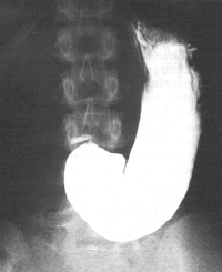 |
Fig. 26.1.
Case G.C. Moderate dilatation of stomach, sagging of greater curvature, and
absence of peristalsis and cyclical activity of pyloric sphincteric cylinder
|
According to Monges et al. (l974) the first electromyographic phenomena of nausea are
the disappearance of spiking activity in the stomach and small intestine, and the slowing,
decrease in amplitude and disturbance in propagation of the basal electrical rhythm
(BER). These events may occur before the patient experiences nausea.
You et al. (l980) recorded gastric myoelectric activity in control subjects and in a group
of patients with unexplained nausea, epigastric bloating and vomiting; the technique
entailed the use of a peroral suction electrode. As no mention was made of actual
retching or vomiting during the investigations, it would appear that recordings were
obtained during the phase of nausea. One of the electrodes was attached to the gastric
mucosa 1.0 cm orally to the pyloric ring, and another 3.0 cm orally to the ring, i.e. within
the confines of the pyloric sphincteric cylinder (others were attached further orally, and 2
were located in the duodenum). In contrast to the normal, regular pacesetter potential
(PP) with a frequency of 3 to 4 cycles per minute, all 9 patients showed abnormal "antral"
myoelectric activity, characterized by tachygastria or tachyarrhythmia and propagation of
the PP in either the orad or aborad direction.
Previous Page | Table of Contents | Next Page
© Copyright PLiG 1998