


Go to chapter: 1 | 2 | 3 | 4 | 5 | 6 | 7 | 8 | 9 | 10 | 11 | 12 | 13 | 14 | 15 | 16 | 17 | 18 | 19 | 20 | 21 | 22 | 23 | 24 | 25 | 26 | 27 | 28 | 29 | 30 | 31 | 32 | 33 | 34 | 35 | 36 | 37 | 38 | 39
Chapter 10 (page 40)

Go to chapter: 1 | 2 | 3 | 4 | 5 | 6 | 7 | 8 | 9 | 10 | 11 | 12 | 13 | 14 | 15 | 16 | 17 | 18 | 19 | 20 | 21 | 22 | 23 | 24 | 25 | 26 | 27 | 28 | 29 | 30 | 31 | 32 | 33 | 34 | 35 | 36 | 37 | 38 | 39
Chapter 10 (page 40)
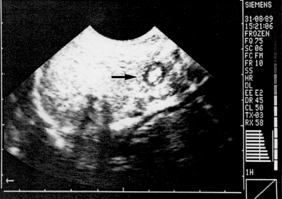 | Fig. 10.1. Transverse ultrasonic section of normal pyloric ring showing "doughnut" (arrow). The hypoechoic ring is the muscular, and the inner echogenic core the mucosal/submucosal component of the ring |
Strauss et al. (l98l), using a static gray-scale B-scan unit and subsequently a real-time unit
with a 5MHz focused transducer, considered the infantile pylorus to be within normal
limits if its overall diameter measured 1.5cm or less.
Longitudinal sections of the normal pylorus, on which the canal length can be measured,
may also be obtained. In tracing the thin, hypoechoic muscular layer distally to the
gastric outlet, Blumhagen and Noble (l983) found the "antral" muscular layer to vary
from 1.5 to 3.0 mm in thickness in normal infants. Khamapirad and Athey (l983), using
digital gray-scale static equipment and a 5MHz focused transducer, studied transverse
and longitudinal sonographic images of the pylorus in 12 normal infants between the ages
of one and 6 weeks. The normal pyloric ring was similar in appearance to the mass of
IHPS but was less than 1.0cm in diameter.
In a control group of 24 normal infants ranging in age from 2 days to 32 weeks,
Graif et al. (l984) found the mean and standard deviation for the transverse diameter to be
7.45 ± 2.2 mm. The mean single wall thickness was 2.3 mm with a standard deviation of
± 0.7 mm, while the mean length of the pylorus was 12.0 mm with a standard deviation
of ± 3.7 mm.
Wilson and Vanhoutte (l984) measured what they called the true pyloric muscle length in
l7 normal babies, and found the range to vary from 12.0 to 15.0 mm.
Stunden et al. (l986), in 88 normal infants under the age of 5 months, found the mean
overall diameter of the pylorus to be 9.1 ± 1.1mm, the mean muscle thickness 1.6 ± 0.4
mm, and the mean canal length 8.3 ± 2.5 mm. These measurements were all obtained
with the pylorus in its most contracted state. Additional information could be obtained
when the pylorus was viewed in real-time. Normally the pyloric "canal" was seen to
relax, allowing fluid to pass from stomach to duodenum. Some variation in overall
diameter and muscle thickness did occur, probably representing alterations in muscle
tone.
According to Stringer et al. (l986) the thickness of the inner echogenic layer, consisting
of the mucosa, muscularis mucosae and submucosa, normally varies between 2.5 and 3.5
mm in infants.
Swischuk (l989) found the hypoechoic outer muscular layer to measure only 1.0 mm in
normal infants. At times it was too thin to be measureable.
Previous Page | Table of Contents | Next Page
© Copyright PLiG 1998