


Go to chapter: 1 | 2 | 3 | 4 | 5 | 6 | 7 | 8 | 9 | 10 | 11 | 12 | 13 | 14 | 15 | 16 | 17 | 18 | 19 | 20 | 21 | 22 | 23 | 24 | 25 | 26 | 27 | 28 | 29 | 30 | 31 | 32 | 33 | 34 | 35 | 36 | 37 | 38 | 39
Chapter 33 (page 167)
Case 33.7 M.B., 68 year old female, presented with a large, hard epigastric mass.
Radiology showed a constant narrowing of the distal 3.0 to 4.0 cm of the stomach,
somewhat resembling partial contraction or spasm of the pyloric sphincteric cylinder
(Fig. 33.7A). However, it contained a filling defect and mucosal folds were
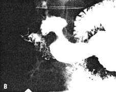
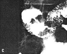
absent. While some degree of movement was seen, this was atypical, with total absence
of cylical contraction and relaxation of the cylinder (Fig. 33.7B - E). The base
of the duodenal bulb appeared normal. Emptying of barium suspension was not
significantly delayed.
At Billroth II partial gastrectomy a large, ulcerated pyloric carcinoma, extending aborally
as far as the pyloric ring, was removed. The duodenum appeared normal. Microscopy
revealed a signet ring cell carcinoma extending as far as the lamina propria and not
involving the muscularis. The duodenum was confirmed to be normal.
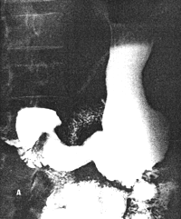 |
Fig. 33.7. A Case M.B. Narrowing distal 3.0 to 4.0 cm of stomach
resembling partial contraction or spasm of sphincteric cylinder.
|
 |  |
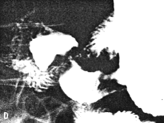 | 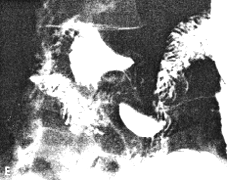 |
| Fig. 33.7 B-E. Case M.B. Filling defect in narrowed region. Mucosal
folds absent. Some movement evident but cyclical contraction and relaxation of
sphincteric cylinder absent. Base of duodenal bulb appears normal.
|
Radiographic Anatomy of Sphincteric Cylinder
In all 50 verified cases of pyloric carcinoma, alteration, deformity or destruction of the
anatomical constituents of the pyloric sphincteric cylinder occurred to greater or lesser
extent. In most cases the cylinder was totally unrecognizable; in 3 an appearance
vaguely simulating the normal cylinder was seen (Fig. 33.7A). In some
cases the process was of a mainly proliferative type, causing intraluminal filling defects
(Fig. 33.1). In others it was mainly infiltrative, causing rigidity of the walls and "fixing"
of the pyloric aperture in the patent or open position, with apparently a normal rate of
emptying of fluid barium (Fig. 33.5). The process was of a mainly stenosing nature in
other cases, with irregularity of the walls and narrowing of the lumen and pyloric
aperture, causing partial or total obstruction (Fig. 33.2, 33.6). In many cases the tumor
mass was ulcerated; not infrequently a combination of the above appearances was seen.
In the few cases where the process somewhat resembled partial contraction of the normal
cylinder, associated filling defects, mucosal destruction and atypical, restricted
movements confirmed the diagnosis.
Of importance is the fact that the narrowing did not tally with any phase of the normal,
cyclical contraction of the sphincteric cylinder.
There was destruction of mucosal folds within the confines of the lesion in all cases.
Motility of Sphincteric Cylinder
In all cases movements of the pyloric sphincteric cylinder were abolished or markedly
altered. Only in 3 cases some movement occurred, but it was atypical (Fig.
33.7B); normal cyclical contraction and relaxation of the cylinder was
absent in all.
Emptying of Liquid Barium
In most cases there was no appreciable delay in the emptying of barium suspension; in a
few cases incomplete or almost complete obstruction to the flow of liquid barium
occurred.
Previous Page | Table of Contents | Next Page
© Copyright PLiG 1998


