

Go to chapter: 1 | 2 | 3 | 4 | 5 | 6 | 7 | 8 | 9 | 10 | 11 | 12 | 13 | 14 | 15 | 16 | 17 | 18 | 19 | 20 | 21 | 22 | 23 | 24 | 25 | 26 | 27 | 28 | 29 | 30 | 31 | 32 | 33 | 34 | 35 | 36 | 37 | 38 | 39
Chapter 33 (page 166)


Go to chapter: 1 | 2 | 3 | 4 | 5 | 6 | 7 | 8 | 9 | 10 | 11 | 12 | 13 | 14 | 15 | 16 | 17 | 18 | 19 | 20 | 21 | 22 | 23 | 24 | 25 | 26 | 27 | 28 | 29 | 30 | 31 | 32 | 33 | 34 | 35 | 36 | 37 | 38 | 39
Chapter 33 (page 166)
 |
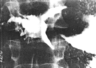
Fig. 33.4.Case A.S. Irregular filling defect distal stomach. Mucosal folds and cyclical activity of cylinder absent. Pyloric aperture unrecognizable. Concave indentation and irregularity of duodenal bulb. |
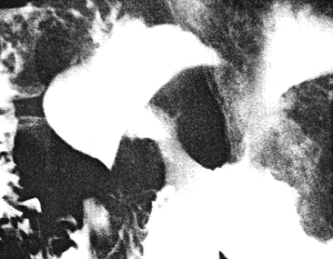
Case 33.5 R.B., 60 year old female, presented with malaena of 5 months' duration,
iron deficiency anaemia and an epigastric mass. Radiology showed a constant, lobulated
and constricting filling defect in the distal 8.0 cm of the stomach, extending as far as the
pyloric aperture, which remained patent. Emptying of fluid barium was not significantly
delayed. Mucosal folds in the affected region and cyclical activity of the pyloric
sphincteric cylinder were absent. The base of the duodenal bulb showed a smooth,
regular, concave indentation, suggestive of external pressure rather than infiltration of the
bulb itself (Fig. 33.5).
Gastroscopy revealed a large ulcerating carcinoma extending to the gastro-oesophageal
junction on the lesser curvature. Endoscopic biopsy showed a poorly differentiated
papillary adenocarcinoma. At laparotomy a large gastric carcinoma was found; there
was infiltration of the transverse colon and gall bladder, with metastases in the liver. It
was difficult to evaluate the duodenum precisely. The tumor was considered to be
unresectable and palliative gastro-enterostomy was done.
 |
Fig. 33.5.Case R.B. Lobulated and constricting filling defect distal stomach. Mucosal folds and cyclical activity of cylinder absent. Pyloric aperture patent. Smooth, concave indentation base of duodenal bulb. |
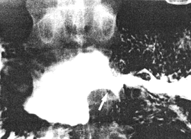
Case 33.6 S.M., 49 year old male, presented with dyspepsia, loss of weight and
postprandial vomiting of 7 months' duration. Radiology showed a constant, irregular
narrowing with nodular filling defects in the distal 5.0 to 6.0 cm of the stomach; aborally
it extended to the pyloric ring. Mucosal folds were not recognizable in the affected
region. There was total absence of cyclical activity of the sphincteric cylinder. Emptying
of fluid barium was not significantly delayed. A concave indentation of the base of the
duodenal bulb, presumably due to external pressure, was seen (Fig. 33.6). In other
respects the bulb appeared normal; the duodenal "tail" was normal.
Gastroscopy showed an infiltrating carcinoma of the "antrum", involving the lesser and
greater curvatures and extending to within 8.0 cm of the gastro-oesophageal junction; the
pyloric aperture could not be visualized. At Billroth II partial gastrectomy the tumor was
removed; it extended to the pylorus but not into the duodenum. The transverse
mesocolon was attached to it; there were no lymphatic or liver metastases. Microscopic
examination revealed a poorly differentiated adenocarcinoma infiltrating deeply into the
muscularis externa. No duodenal extension was seen. Draining lymph glands were
normal. The surrounding gastric mucosa showed subacute gastritis.
 |
Fig. 33.6. Case S.M. Irregular narrowing distal stomach. Mucosal folds and cyclical activity of cylinder absent. Pyloric aperture deformed. Concave indentation base of duodenal bulb. Duodenal "tail" (arrow) apparently unaffected. |
Previous Page | Table of Contents | Next Page
© Copyright PLiG 1998