


Go to chapter: 1 | 2 | 3 | 4 | 5 | 6 | 7 | 8 | 9 | 10 | 11 | 12 | 13 | 14 | 15 | 16 | 17 | 18 | 19 | 20 | 21 | 22 | 23 | 24 | 25 | 26 | 27 | 28 | 29 | 30 | 31 | 32 | 33 | 34 | 35 | 36 | 37 | 38 | 39
Chapter 33 (page 169)

Go to chapter: 1 | 2 | 3 | 4 | 5 | 6 | 7 | 8 | 9 | 10 | 11 | 12 | 13 | 14 | 15 | 16 | 17 | 18 | 19 | 20 | 21 | 22 | 23 | 24 | 25 | 26 | 27 | 28 | 29 | 30 | 31 | 32 | 33 | 34 | 35 | 36 | 37 | 38 | 39
Chapter 33 (page 169)
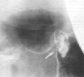 |
Fig. 33.9. Case A.F. Small mass lesion at pyloric aperture (arrow). Base of gas-filled duodenal bulb normal. |
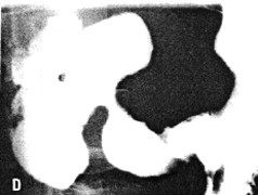
Case 33.10 S.F., 49 year old male. Radiology: 6.0 cm nodular and constricting
pyloric filling defect. Smooth, concave indentation base of duodenal bulb (Fig. 33.10).
Operation: Pyloric mass with serosal spread, adherent to pancreas. Lymph node
metastases. Billroth II. Gastric histology: well differentiated adenocarcinoma. Duodenal
histology: spread into serosa and muscularis of duodenum. Duodenal mucosa and
Brunner's glands free of tumor cells.
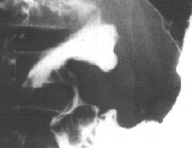 |
Fig. 33.10.Case S.F. Nodular and constricting pyloric filling defect. Smooth, concave indentation base of duodenal bulb. |
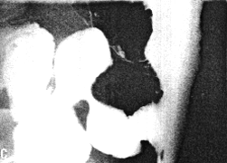
Case 33.11 K.B., 69 year old female. Radiology: 6.0 cm long constricting pyloric
filling defect. Base of duodenal bulb normal (Fig. 33.11). Operation: Pyloric mass with
serosal spread and lymph node metastases. Billroth II. Gastric histology: poorly
differentiated adenocarcinoma. Marked infiltration of pyloric ring region. Duodenal
histology: some tumor cells in muscularis and small blood vessels. Brunner's glands not
affected.
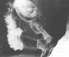 |
Fig. 33.11.Case K.B. Constricting pyloric filling defect. Base of duodenal bulb normal. |
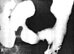
Case 33.12 E.L.J., 44 year old male. Radiology: 7.0 cm long constricting pyloric
filling defect. Base of duodenal bulb normal (Fig. 33.12). Operation: large mass pyloric
region with serosal spread. Duodenum appears normal. Widespread lymphatic
metastases. Coeliac and para-aortic glands involved. Metastases in transverse
mesocolon and spleen. Billroth II. Gastric histology: poorly differentiated
adenocarcinoma (mucinous type with signet ring cells). Duodenal histology: spread into
submucosa up to commencement of Brunner's glands. No infiltration of Brunner's glands
or superficial mucosa.
 |  |
 |  |
| Fig. 33.12 A-D Case E.L.J. Constricting pyloric filling defect. Base of duodenal bulb normal. | |
Previous Page | Table of Contents | Next Page
© Copyright PLiG 1998