

Go to chapter: 1 | 2 | 3 | 4 | 5 | 6 | 7 | 8 | 9 | 10 | 11 | 12 | 13 | 14 | 15 | 16 | 17 | 18 | 19 | 20 | 21 | 22 | 23 | 24 | 25 | 26 | 27 | 28 | 29 | 30 | 31 | 32 | 33 | 34 | 35 | 36 | 37 | 38 | 39
Chapter 3 (page 13)


Go to chapter: 1 | 2 | 3 | 4 | 5 | 6 | 7 | 8 | 9 | 10 | 11 | 12 | 13 | 14 | 15 | 16 | 17 | 18 | 19 | 20 | 21 | 22 | 23 | 24 | 25 | 26 | 27 | 28 | 29 | 30 | 31 | 32 | 33 | 34 | 35 | 36 | 37 | 38 | 39
Chapter 3 (page 13)
 | Fig. 3.4. F.M., fan-shaped muscle according to Cole; its concentric contraction causes formation of the pyloric canal; P.A., pyloric aperture |
Torgersen (l942) studied the muscular build and movements of the stomach and duodenal
bulb from the point of view of comparative anatomy and embryology. Although his
methodology was quite different from that of Forssell (1913), his results verified the
latter's conception of the canalis egestorius in all important respects and he accepted
Forssell's terminology. He differed from Forssell in a few details; for instance, whereas
Forssell included part of the membrana angularis on the lesser curvature in the canalis
egestorius, Torgersen regarded these as two separate regions.
Torgersen (l942) was able to add important new findings which further elucidated the
muscular anatomy of the "transverse" part of the stomach. His monumental work
commenced with an historical review of the anatomy of the stomach from the time of
Willis (1682) to the era following Forssell (1913). He showed in detail how previous
anatomists such as Retzius, Luschka, von Aufschnaiter, Jonnesco and E. Müller opened
the way for Cunningham (l906) and Forssell (l913). On the other hand a few anatomists,
the most notable being Pernkopf (l922, l924), differed from the latter; while Forssell held
that the musculature of the stomach was highly differentiated into separate but contiguous
regions, Pernkopf maintained that there was no differentiation in the musculature at all.
According to Pernkopf the regions lacked anatomical foundation, and the forms of
movement were entirely of a functional nature; nevertheless he agreed that the
movements were not devoid of comparative anatomical interest, as they imitated the
more complex stomachs of other vertebrates.
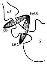
According to Torgersen (l942) the circular musculature of the canalis egestorius in man
and other vertebrates contains two annular thickenings or loops. The aboral loop is called
the right canalis loop (Fig. 3.5). (Comment: At times he also referred to this loop as the
pyloric sphincter; the word "sphincter" was an unfortunate choice, as it will become clear
that Torgersen regarded the "sphincter" as a complex structure consisting of various
loops, of which the right canalis loop constituted but one component. In a personal
communication to the present author in 1962, Torgersen confirmed that the right canalis
loop was the muscular component of the pyloric ring).
 | Fig. 3.5. Diagram of circular musculature of sphincteric cylinder (canalis egestorius) according to Torgersen. R.P.L., right pyloric (canalis) loop; L.P.L., left pyloric (canalis) loop; P.M.K., pyloric muscle knot (torus); S, stomach; D.B., duodenal bulb. (Ring of circular musculature surrounding commencement of duodenum not shown.) |
Previous Page | Table of Contents | Next Page
© Copyright PLiG 1998