


Go to chapter: 1 | 2 | 3 | 4 | 5 | 6 | 7 | 8 | 9 | 10 | 11 | 12 | 13 | 14 | 15 | 16 | 17 | 18 | 19 | 20 | 21 | 22 | 23 | 24 | 25 | 26 | 27 | 28 | 29 | 30 | 31 | 32 | 33 | 34 | 35 | 36 | 37 | 38 | 39
Chapter 3 (page 14)
The left canalis loop is located on the oral side of the right loop. The two loops, each
being placed obliquely, meet on the lesser curvature in a muscle torus (Fig. 3.5). From
the torus the loops diverge to encircle the greater curvature, where they are 3.0 to 5.0 cm
apart. It is evident from the course of the fibres that the two loops are not independent
anatomical structures; their musculature is intimately interlaced in the muscle torus on
the lesser curvature, and also with the intervening circular muscle fibres in the anterior
and posterior gastric walls.
The left canalis loop corresponds to the sulcus intermedius on the greater curvature. The
circular fibres on the oral side of the left loop merge imperceptibly into the circular fibres
of the adjacent sinus. The circular musculature of the canalis is thicker than that of the
sinus, but in other respects no boundary can be demonstrated between these two regions.
On the lesser curvature the concentrated circular musculature in the muscle torus is
continuous with the thin musculature of the membrana angularis.
Torgersen (l942) found that the two loops were distinctly visible in some of the
illustrations presented by previous anatomists such as Cunningham (1906), Wernstedt
(who named the left loop the sphincter intermedius), and even Pernkopf (l921, l924).
On the duodenal side a connective tissue septum separates the main mass of the right
canalis loop from the circular fibres of the duodenum. On the aboral side of the septum a
strong loop of circular musculature surrounds the base of the duodenal bulb. A few of
the circular fibres of the muscle torus on the lesser curvature are continuous with those of
the duodenal loop. In the anterior and posterior gastric walls the right canalis loop and
circular duodenal loop are loosely connected by the intervening connective tissue septum
and a few muscular anastomoses. On the greater curvature the right canalis loop is
connected more intimately to the circular duodenal loop.
Torgersen (l942) regarded the circular muscle loop at the base of the duodenal bulb as
part of the pyloric sphincteric mechanism. In his view the pyloric sphincter, as far as the
circular musculature was concerned, consisted of gastric and duodenal parts, viz. the right
and left canalis loops on the gastric side, and the loop surrounding the commencement of
the duodenum on the aboral side of the fibrous tissue septum.
The longitudinal musculature of the sinus becomes abruptly thicker at the left canalis
loop and forms a powerful band between the right and left loops on the greater curvature,
according to Torgersen (l942). The majority of these longitudinal fibres, as well as those
in the anterior and posterior gastric walls, dip into the musculature of the right canalis
loop (i.e. the muscular component of the pyloric ring); only a few longitudinal fibres are
carried across the connective tissue septum into the duodenum on the greater curvature
side. On the lesser curvature side most longitudinal bundles proceed uninterruptedly
across the septum into the duodenum.
According to Torgersen (l942) the canalis egestorius consists of the muscle torus, the left
and right circular loops, and the circular and longitudinal muscle fibres between these
structures. The sphincteric mechanism at the pylorus consists of the canalis egestorius,
the circular musculature surrounding the commencement of the duodenum, and the
intervening fibrous septum.
Torgersen (l942) found the muscular build of the stomach and duodenum in the dog,
rabbit, horse, pig and ox to be essentially similar to that in man. In all these animals the
right and left circular loops, the muscle torus on the lesser curvature and the longitudinal
fibres between the circular loops were clearly discernible. The circular fibres at the
commencement of the duodenum, the connective tissue septum between these and the
right loop, and the arrangement of the longitudinal fibres across the septum were also
similar to that in man. There were a few minor variations; for instance, the circular
canalis loops in the dog appeared to be more powerful on the greater curvature side,
while in the horse the loops were equally powerful throughout their circumference. The
duodenal bulb was more prominent in man and the horse than in the other animals
studied. In the cat the right loop differed in that it was only developed on the greater
curvature side.
Torgersen (l942) concluded that there was a common principle of build of this part of the
stomach in the higher vertebrates and in man. He found the canalis to be an anatomically
preformed structure, an anatomical reality with a sound foundation in comparative
anatomy.
McNaught (l957) confirmed the presence of the left canalis loop in fresh gastric resection
specimens. Williams (l962) stated that the contracted fan-shaped muscle was
occasionally seen in fresh partial gastrectomy specimens.
Keet and Heydenrych (l982) studied the width of the gastric walls in the pyloric region in
adults, in 5 morbid anatomical specimens fixed in formalin. Having identified the pyloric
ring by means of a wire marker, the lumen of the stomach and duodenum was filled with
barium. A narrow layer of barium paste was painted on the serosal surface of the lesser
curvature, and another on the greater curvature. Radiographs of each specimen were
taken in the anteroposterior position (Fig. 3.6). The space between the luminal barium
and that on the external surface indicated the thickness of the wall, consisting of mucosa,
submucosa, muscularis externa and serosa. As the mucosal, submucosal and serosal
layers were more or less uniformly thick in all parts of the stomach, any variation in wall
thickness would be due to thickening of the muscular coat.
In all specimens the following was seen: extending orally from the pyloric ring there was
a cylindrical region approximately 3.0 cm in length in which the wall had a thickness of
6.0 to 7.0 mm; it was slightly shorter on the lesser than on the greater curvature. In the
remainder of the stomach the wall thickness was 2.0 to 3.0 mm. The pyloric ring formed
the aboral part of the muscular thickening.
It was concluded that there was a tube of thickened pyloric musculature, approximately
3.0 cm in length and incorporating the pyloric ring, in adult morbid anatomical
specimens.
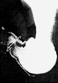 | Fig. 3.6.
Radiograph of morbid anatomical specimen. Barium fills the lumen and outlines the
serosa of the lesser and greater curvatures. A short tube of thickened muscularis
externa extends orally from the pyloric ring
|
Previous Page | Table of Contents | Next Page
© Copyright PLiG 1998