


Go to chapter: 1 | 2 | 3 | 4 | 5 | 6 | 7 | 8 | 9 | 10 | 11 | 12 | 13 | 14 | 15 | 16 | 17 | 18 | 19 | 20 | 21 | 22 | 23 | 24 | 25 | 26 | 27 | 28 | 29 | 30 | 31 | 32 | 33 | 34 | 35 | 36 | 37 | 38 | 39
Chapter 28 (page 135)


Go to chapter: 1 | 2 | 3 | 4 | 5 | 6 | 7 | 8 | 9 | 10 | 11 | 12 | 13 | 14 | 15 | 16 | 17 | 18 | 19 | 20 | 21 | 22 | 23 | 24 | 25 | 26 | 27 | 28 | 29 | 30 | 31 | 32 | 33 | 34 | 35 | 36 | 37 | 38 | 39
Chapter 28 (page 135)
 |  |
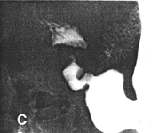 | 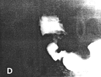 |
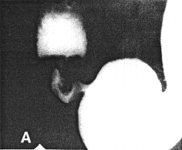
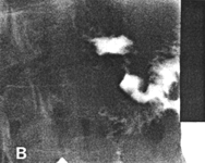
| Fig. 28.2 A-D. Case J.C. Constant contraction of sphincteric cylinder with absent cyclical activity. Irregular, immobile folds in cylinder | |
Peristaltic waves were normal in the remainder of the stomach, but stopped abruptly at
the commencement of the cylinder, which showed total absence of cyclical contraction
and relaxation. Endoscopy revealed chronic "antral" gastritis; a few small erosions were
noted in the first part of the duodenum.
Case 28.3 L.M., 50 year old female. Radiographic examination showed pronounced,
constant contraction of the pyloric sphincteric cylinder; occasionally a minor degree of
relaxation occurred, but cyclical contraction and relaxation was absent and most of the
time the appearance remained as illustrated (Fig. 28.3). Prominent circular and irregular
mucosal folds which appreared to be immobile, were present in the contracted cylinder.
A concave indentation was seen in the base of the duodenal bulb. Endoscopy revealed a
number of mucosal erosions as well as an appearance of severe "antral" gastritis. This
was still present at a second endoscopy 8 months later, indicating chronicity.
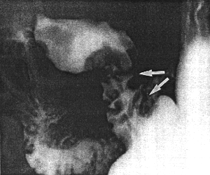 |
Fig. 28.3. Case L.M. Constant contraction of sphincteric cylinder (arrows), with prominent irregular, static mucosal folds |
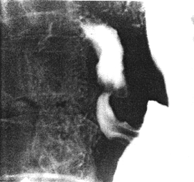
Case 28.4 C.C., 36 year old female. Radiographic examination showed the pyloric sphincteric cylinder to be markedly contracted most of the time; at such times it contained longitudinal mucosal folds (Fig. 28.4A). Occasionally it relaxed to a certain extent, when a fold changed in direction to become circular (Fig. 28.4B); relaxation was never complete and cyclical activity was absent. Endoscopy revealed an erosion on the lesser curvature side of the pyloric region; touching the mucosa caused haemorrhage. The endoscopic diagnosis was erosive antral gastritis.
 A A | |
| Fig. 28.4. A Case C.C. Marked contraction of sphincteric cylinder, with longitudinal mucosal folds B Case C.C. Some relaxation of cylinder. A mucosal fold has changed in direction to become circular. Cyclical activity absent | 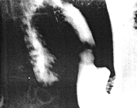 B B |
Previous Page | Table of Contents | Next Page
© Copyright PLiG 1998