

Go to chapter: 1 | 2 | 3 | 4 | 5 | 6 | 7 | 8 | 9 | 10 | 11 | 12 | 13 | 14 | 15 | 16 | 17 | 18 | 19 | 20 | 21 | 22 | 23 | 24 | 25 | 26 | 27 | 28 | 29 | 30 | 31 | 32 | 33 | 34 | 35 | 36 | 37 | 38 | 39
Chapter 28 (page 136)
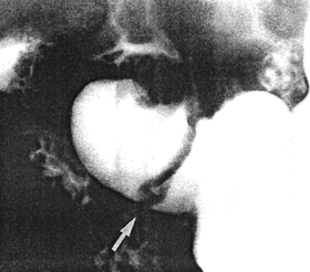
Case 28.5 J.M., 45 year old male. The pyloric sphincteric cylinder remained
partially contracted throughout the radiographic examination, with complete absence of
normal, cyclical contraction and relaxation. At least one prominent, circumferential
mucosal fold, which did not change in position, was present in the partially contracted
cylinder (Fig. 28.5). Initially it was difficult to distinguish between a permanent,
circumferential mucosal fold and a prepyloric septum. Endoscopy revealed prominent,
thickened prepyloric mucosal folds with an abnormal, whitish, granular surface.
Endoscopic biopsy showed lymphocytic and plasma cell infiltration of the lamina
propria; in some areas atypical epithelium, presumably due to inflmmatory change, was
seen. The endoscopic diagnosis was chronic "antral" gastritis.
 |
Fig. 28.5.
Case J.M. Partial contraction of sphincteric cylinder, with absent cyclical activity.
Prominent static, circular mucosal fold in cylinder (arrow)
|
A brief review of the literature shows that many aspects of gastritis are still controversial;
even its definition is contentious. It has been stated that clinicians, pathologists and
endoscopists defined "gastritis" in different ways (Hojgaard et al. l987). Others held that
the term "chronic gastritis" had different connotations for the clinician, the pathologist
and radiologist (Rao et al. l975). Authors differ on the subdivision of gastritis into
various types and grades. It is agreed, however, that the diagnosis in the intact stomach
can only be made by means of endoscopy, biopsy and microscopy. As the deeper layers
of the wall are also involved in many instances, a full investigation would entail
histologic examination of resection specimens.
The cases quoted here show that certain radiologically recognizable alterations may be
associated with chronic "antral" gastritis, perhaps more correctly termed chronic gastritis
affecting the pyloric sphincteric cylinder. (One of the cases was diagnosed as erosive
haemorrhagic gastritis). The radiologically recognizable alterations are:
- Partial but constant contraction of the sphincteric cylinder; the degree of
contraction was usually described as "moderate" or "marked". In these cases
radiologically visible peristaltic waves in the corpus and sinus of the stomach
appeared normal, but each wave stopped at the commencement of the partially
contracted cylinder. In some cases the cylinder showed minor degrees of
contraction and relaxation, but maximal cyclical contraction and relaxation at a
rate of 3 cycles per minute was absent in all. The appearance closely resembles
that of pylorospasm (Chap. 20).
In some cases the pyloric aperture was seen to be fixed in the open position as a
consequence of partial contraction of the cylinder; this appearance may be
associated with duodenogastric reflux (Chap. 27). It is presumed that the lack of
cyclical activity of the cylinder may have a bearing on trituration and the
emptying of solids (Chap. 18).
- Prominent irregular and/or circular mucosal folds showing restricted movements
or no movement at all. In view of the restricted movements of the walls of the
cylinder it is not surprising that "independent but co-ordinated" movements of the
inner mucosal layer should also be curtailed or absent (Chaps. 2, 13). It is
surmized that cellular infiltration in the mucosa, submucosa, muscular layers and
neuronal elements occurring in chronic gastritis, partially accounts for the
restricted movements.
Theoretically the static, irregular mucosal folds projecting into the lumen should
hamper duodenogastric reflux occurring as a result of patency of the pyloric
aperture in some cases.
- A presumptive radiological diagnosis of chronic gastritis affecting the sphincteric
cylinder, was only made if the above features occurred in the absence of other
pathology in the upper gastrointestinal tract (e.g. gastric ulceration or hiatus
hernia).
According to Nesland and Berstad (l985) and Karvonen et al. (l987), erosive
prepyloric change (EPC) was a condition characterized by standing mucosal folds
and erosions; it was diagnosed endoscopically and appeared to be an entity of its
own. Radiographically a standing mucosal fold may also be recognized, as seen
in Fig. 28.5. In this case it concerned a circular fold which failed to change in
position. It was associated with lymphocytic and plasma cell infiltration, no
mention being made of erosions.
Previous Page | Table of Contents | Next Page
© Copyright PLiG 1998


