

Go to chapter: 1 | 2 | 3 | 4 | 5 | 6 | 7 | 8 | 9 | 10 | 11 | 12 | 13 | 14 | 15 | 16 | 17 | 18 | 19 | 20 | 21 | 22 | 23 | 24 | 25 | 26 | 27 | 28 | 29 | 30 | 31 | 32 | 33 | 34 | 35 | 36 | 37 | 38 | 39
Chapter 28 (page 134)
Nesland and Berstad (l985) noted a specific endoscopic appearance, consisting of
standing prepyloric mucosal folds, redness and erosions, in a considerable number of
patients presenting with dyspepsia; they called the condition erosive prepyloric changes
(EPC) and divided it into three grades on the basis of the endoscopic features. In grade 1
there were standing mucosal folds, independent of peristalsis, running either transversely
or longitudinally in the "antral" lumen. In grade 2 the same appearance was seen with red
spots or streaks situated on top of the folds. In grade 3 the previous 2 appearances were
associated with macroscopic erosions, described as white, fibrin-covered spots with red
halos. EPC grades 2 and 3 were found in 26 percent of 1001 consecutive patients
examined endoscopically. After excluding all patients with ulcer, carcinoma, post-
operative conditions and upper gastro-intestinal bleeding, a group of 651 patients with
non-ulcer dyspepsia remained; EPC grades 2 or 3 were seen in 210. In a group of 34
asymptomatic volunteers, grade 2 was found in 5 cases and grade 3 in one case. The
frequency of grades 2 and 3 of erosive prepyloric changes appeared to be higher in non-
ulcer dyspepsia than in patients with peptic ulceration and also varied with age. The
highest frequency encountered was 52 percent, in patients with non-ulcer dyspepsia in the
40 to 49 year age group. The entity prepyloric erosive changes probably represented a
form of antral gastritis. It might have a bearing on the radiological diagnosis of antral
gastritis. Although the redness and often also the erosions were not visible
radiologically, the permanent and coarse mucosal folds should be evident.
Berstad and Nesland (l985) stated that the three grades of EPC were merely different
expressions of the same process. While the superficial mucosal state might regress or
progress to another grade, the prominent, standing prepyloric mucosal folds were of a
permanent nature. Histological verification was obtained in 88 percent of cases of
endoscopically visible grade 3 erosions; they were always accompanied by an element of
acute inflammation with infiltration of neutrophil polymorphs. In the vast majority there
was an additional chronic inflammatory infiltration of lymphocytes and plasma cells.
Intramucosal fibrosis was present in all biopsy specimens. Clinically, EPC appeared to
be related to non-ulcer dyspepsia. The symptoms could be suggestive of gastric ulcer,
while endoscopy proved the absence of ulceration. The findings supported the theory
that EPC was a disease entity of its own, to be differentiated from ulcer disease. The
permanent feature of the condition, namely standing mucosal folds, was independent of
the ongoing inflammatory activity in the surface layer; whether contractions in the
deeper muscular layers contributed to the fold formation was not known.
Berstad and Nesland (l987) reiterated that EPC was an endoscopic diagnosis based on the
presence of standing prepyloric folds, with or without different types of erosions.
Standing folds were defined as transverse or parallel folds in the prepyloric region, which
might run into the pylorus itself and which were independent of peristaltic movements.
They did not believe that EPC and peptic ulceration were aspects of the same disease
process; EPC could not be considered to be merely a form of antral gastritis.
Hojgaard et al. (l987) pointed out that clinicians, endoscopists and pathologists defined
gastritis in different ways. Pathoanatomical gastritis occurred very commonly and the
prevalence increased with age. These authors found no correlation between dyspepsia (in
patients without peptic ulceration) and endoscopic signs suggesting gastritis, or
histological gastritis. It was concluded that gastritis did not seem to constitute a clinical
entity in non-ulcer dyspepsia.
Karvonen et al. (l987) studied 130 patients with gastric mucosal erosions, occurring in
the absence of peptic ulceration, by endoscopic biopsy; most were located in the
"antrum". In their view erosions were inconsistent phenomena, probably with different
pathogeneses and etiologies. Some incomplete erosions were associated with abuse of
analgesics, but those located on prepyloric mucosal folds appeared to be associated with
duodenal ulceration or duodenitis. The study suggested that prepyloric erosions
constituted an entity of their own; in most instances the mucosa of the corpus was well
preserved despite ageing, the appearance being similar to that of duodenal ulcer disease.
During routine upper gastrointestinal barium examinations, a presumptive radiological
diagnosis of "antral gastritis" was made from time to time. Most of these patients
subsequently had endoscopic investigations (unfortunately biopsies were not taken in all).
After a suitable interval the patients' clinical notes were perused and a final diagnosis
obtained.
A total of 50 cases who were diagnosed radiologically, and confirmed endoscopically, as
chronic "antral" gastritis were studied. The following are examples of the more
pronounced cases:
Case Reports
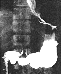
Case 28.1. G.G., 52 year old male. Radiographic examination showed a moderate
degree of constant contraction of the pyloric sphincteric cylinder, with absence of normal,
cyclical contraction and relaxation; the contraction fixed the pyloric aperture in the open
position (Fig. 28.1). The stomach appeared to be hypertonic; rapid emptying of fluid
barium occurred. Endoscopic biopsy of the "antral" region revealed acute on chronic
gastritis; no evidence of malignancy was seen. Repeat endoscopic biopsy two months
later showed acute on chronic inflammatory reaction in the lamina propria, with
eosinophylic infiltration and without evidence of intestinal metaplasia. Stainings for
Helicobacter pylori were positive. A third endoscopic biopsy two months after the
second, showed chronic gastritis with intestinal metaplasia. Stainings for Helicobacter
pylori were negative.
 |
Fig. 28.1.
Case G.G. Moderate degree of constant contraction of pyloric sphincteric
cylinder (arrows). Cyclical activity absent. Pyloric aperture patent
|
Previous Page | Table of Contents | Next Page
© Copyright PLiG 1998


