


Go to chapter: 1 | 2 | 3 | 4 | 5 | 6 | 7 | 8 | 9 | 10 | 11 | 12 | 13 | 14 | 15 | 16 | 17 | 18 | 19 | 20 | 21 | 22 | 23 | 24 | 25 | 26 | 27 | 28 | 29 | 30 | 31 | 32 | 33 | 34 | 35 | 36 | 37 | 38 | 39
Chapter 32 (page 159)
Because of the high incidence of a contracted prepyloric segment in cases of hiatus hernia
in infants as reported by Roviralta (l95l), Astley and Carr‚ (l954), Forshall (l955) and
Johnston (l960), and in adults as reported by Wieser et al. (l963) and Keet and
Heydenrych (l97l), we have to agree with Astley and Carr‚ (l954) that the possibility of a
causal relationship exists. Consequently an investigation was instituted to study the
changes in the pyloric part of the stomach (if any), brought about by electrical and
mechanical stimulation of the structures in and surrounding the oesophageal hiatus in the
diaphragm, including the anterior and posterior vagus nerves and the intrahiatal part of
the oesophagus.
Having received approval of the Ethical Committee, laparotomy was performed on 6
mongrel dogs under general pentothal anaesthesia. No premedication was given, but 50
mg scoline per half hour plus oxygen and a manual respirator were used. The
oesophagus and anterior and posterior vagus nerves were dissected free, as in the
technique used for truncal vagotomy. The electrodes of an A.C. Ruhmkorff induction
coil were attached to various areas, including the anterior and posterior branches of the
vagus. The strength of the induced current was adjustable but the same intensity was
applied to all areas.
In all the experimental animals the changes occurring in the pyloric and duodenal areas
were unequivocal, as follows:
- Stimulation of the tissues surrounding the vagus nerves caused no alteration.
Following stimulation of either the anterior or posterior vagus, at a level just above the
gastroesophageal junction, there was a tubular contraction of the pyloric region of the
stomach, extending orally from and including the pyloric ring. The contracted area
varied in length with the size of the animal, being from 3.0 to 6.0 cm long, and never less
than 3.0 cm (Fig. 32.7A). The elapsed time between stimulation and
contraction was 2 to 3 seconds. The remainder of the stomach remained flaccid. Ceasing
the stimulation caused the contraction to relax.
- The degree of contraction was proportional to the strength of the stimulus. Increasing
the stimulus caused increased contraction of the pyloric segment, until it became a solid
cylinder. The muscular contraction sqeezed out the blood, the firmly contracted region
assuming an anemic, grayish white appearance (Fig. 32.7B). It felt rubbery
hard and resembled a neoplasm. Usually there was some contraction of the first and
second parts of the duodenum as well, but this commenced a second or two later and was
much less marked than the contraction of the pyloric region.
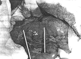
| Fig. 32.7. A
Pyloroduodenal junction in the dog. The area in the pyloric region which will undergo
contraction is demarcated by the limbs of the forceps (retouched) and is 3.0 cm in length.
|
 |
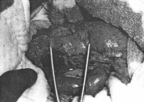 |
Fig. 32.7. B
Following stimulation of the vagus in the hiatus, the pyloric sphincteric cylinder,
demarcated by the forceps, becomes firmly contracted, rubbery hard and anaemic
|
In one of the dogs an attempt was made to simulate the mechanical contraction which is
probably produced on the gastric fornix in cases of hiatus hernia. This was done by
slipping a plastic ball 5.0 cm in diameter into the fornix of the stomach via a gastrostomy.
The walls of the fornix were then compressed manually against the balloon, i.e. the
compressing fingers surrounded the gastric walls, (including the intact vagus nerves), and
compressed these against the balloon in the lumen. A contraction of the pars pylorica,
similar to that described above, occurred.
Previous Page | Table of Contents | Next Page
© Copyright PLiG 1998