

Go to chapter: 1 | 2 | 3 | 4 | 5 | 6 | 7 | 8 | 9 | 10 | 11 | 12 | 13 | 14 | 15 | 16 | 17 | 18 | 19 | 20 | 21 | 22 | 23 | 24 | 25 | 26 | 27 | 28 | 29 | 30 | 31 | 32 | 33 | 34 | 35 | 36 | 37 | 38 | 39
Chapter 31 (page 153)


Go to chapter: 1 | 2 | 3 | 4 | 5 | 6 | 7 | 8 | 9 | 10 | 11 | 12 | 13 | 14 | 15 | 16 | 17 | 18 | 19 | 20 | 21 | 22 | 23 | 24 | 25 | 26 | 27 | 28 | 29 | 30 | 31 | 32 | 33 | 34 | 35 | 36 | 37 | 38 | 39
Chapter 31 (page 153)
 |
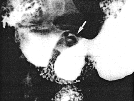
Fig. 31.2. Case J.N. Fistula between pyloric sphincteric cylinder and base of duodenal bulb on lesser curvature side (arrow). Constant contraction of sphincteric cylinder. Pyloric aperture patent |
Anti-ulcer treatment was commenced. Initially the patient was lost to follow-up, but
endoscopy 3 months later showed a posteriorly situated, deep prepyloric ulcer. No
definite penetration into the duodenum could be demonstrated, but the examination was
difficult and incomplete on account of fixation of tissues. After an episode of
haematemesis a month later, a second endoscopy showed the same ulcer to be filled with
blood clot. Because of increasing fixation it was not possible to manipulate the
instrument through the pylorus into the duodenum.
The patient returned a year later after a massive haematemesis. Following ressuscitation,
a laparotomy was performed at which an inflammatory mass was palpated on the
posterior aspect of the pylorus. After a truncal vagotomy had been performed it was
found that a pyloric ulcer had penetrated deeply into the pancreas, necessitating
considerable dissection. The pancreas formed the base of the ulcer; from here it had
burrowed into the duodenum. The pyloric ring was intact. The findings confirmed the
presence of a gastro-duodenal fistula as a result of a penetrating pyloric ulcer. An
antrectomy with Billroth I anastomosis was done. Histologically the ulcer was benign,
with chronic gastritis and intestinal metaplasia in the surrounding gastric mucosa.
Case 31.3 S.D., 44 year old female, was a known case of pyloric ulceration. Three
years before admission an active ulcer on the lesser curvature of the pyloric sphincteric
cylinder had been diagnosed at radiological examination. Follow-up endoscopy after
anti-ulcer therapy had shown healing of the ulcer with some residual deformity of the
pyloric region.
She then presented with a recurrence of symptoms. Endoscopy showed pyloric
deformity. Radiographic examination three weeks later revealed an ulcer on the lesser
curvature of the pyloric sphincteric cylinder, on the immediate oral side of the ring, with
a fistulous communication between the ulcer and the superior fornix of the duodenal bulb
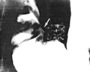
(Fig. 31.3). Prominent, permanent circular mucosal folds were present in the cylinder,
which remained partially expanded throughout the examination with complete absence of
cyclical contraction and relaxation. Repeat endoscopy the following week confirmed the
presence of a prepyloric ulcer with a pyloro-duodenal fistula.
 |
Fig. 31.3. Case S.D. Ulcer lesser curvature side of sphincteric cylinder with fistula (arrow) to base of duodenal bulb. Partial expansion of cylinder. Permanent, circular mucosal folds in cylinder |
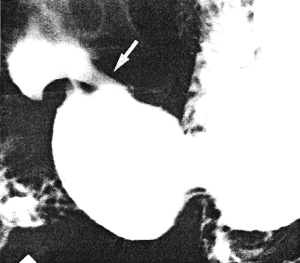
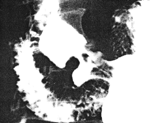
Case 31.4 W.S., 54 year old male, presented with a history of intermittent epigastric pain, aggravated by meals, relieved by alkalies and at times associated with vomiting, of 4 years' duration. Physical examination revealed epigastric tenderness. The first endoscopic examination showed a small, superficial, benign ulcer on the lesser curvature of the stomach approximately l.0cm proximal to the pylorus. After initial improvement the symptoms recurred. Radiographic examination three years later showed a narrow fistula extending from the lesser curvature of the pyloric sphincteric cylinder, 1.0cm proximal to the pylorus, to the superior fornix of the duodenal bulb. The cylinder remained in a state of partial contraction throughout the examination, while the pyloric aperture appeared normal. Control endoscopic examination confirmed the presence of a benign prepyloric ulcer, described as "deep"; the first part of the duodenum was deformed but a pyloro-duodenal fistula could not be identified. A second radiographic study confirmed the findings of the first. At the third radiological study (six months after the first) a fistulous communication was again noted between the prepyloric lesser curvature ulcer and the superior fornix of the duodenal bulb (Fig. 31.4A). The pyloric sphincteric cylinder remained partially contracted throughout the examination, occasionally reaching the pseudo-diverticulum stage, but never relaxed fully (Fig. 31.4B). Subsequently truncal vagotomy and antrectomy was done elsewhere for "chronic prepyloric ulcer". Unfortunately the resection specimen was discarded and was not available for examination.
A | B |
| Fig. 31.4 A,B. Case W.S. A Fistula (arrow) between sphincteric cylinder and duodenal bulb on lesser curvature side. Cylinder partially contracted. Pyloric aperture patent. B Sphincteric cylinder contracted to pseudo-diverticulum stage. It never relaxed fully | |
Previous Page | Table of Contents | Next Page
© Copyright PLiG 1998