

Go to chapter: 1 | 2 | 3 | 4 | 5 | 6 | 7 | 8 | 9 | 10 | 11 | 12 | 13 | 14 | 15 | 16 | 17 | 18 | 19 | 20 | 21 | 22 | 23 | 24 | 25 | 26 | 27 | 28 | 29 | 30 | 31 | 32 | 33 | 34 | 35 | 36 | 37 | 38 | 39
Chapter 31 (page 152)
Chapter 31
Pyloroduodenal Fistula or Acquired Double Pylorus
Whereas a double pylorus had previously been presumed to be of congenital origin,
Hansen et al. (1972) described 2 cases in which the clinical and endoscopic findings
indicated that it was usually an acquired lesion. The first case was a 55 year old male
with a known prepyloric gastric ulcer. Follow-up gastroscopy showed that the ulcer had
perforated into the duodenum, forming a short fistulous communication between the
stomach and duodenum, which had the appearance of a second pyloric aperture. In the
second case a known duodenal ulcer had perforated through the pyloric ring into the
stomach, with a similar result. In both cases a mucosal septum was situated between the
2 apertures.
Drapkin et al. (1974) described a case in which a deep ulcer on the lesser curvature of the
"antrum" eventually perforated into the duodenal bulb, forming an acquired pyloro-
duodenal fistula. At endoscopy two pyloro-duodenal openings, separated by a mucosal
septum, were seen. A catheter inserted into one opening re-entered the "antrum" through
the other.
Engle (1975) collected 7 cases from the literature and described another case in which a
prepyloric gastric ulcer had penetrated into the duodenum. The radiological appearance
was that of a short gastroduodenal fistula, extending from the distal stomach to the
duodenal bulb on the lesser curvature side. The accessory canal was separated from the
normal pylorus by a septum or bridge of mucosa, which conceivably could simulate a
filling defect or mass lesion at a radiographic study. It was stated that cases had probably
been misdiagnosed previously and that the condition was more common than the number
of reported cases would suggest. A similar case was reported by Bender and Soffa
(1975).
Hegedus et al. (1978) studied the developmental history as well as the clinical,
endoscopic and radiological features of 11 cases of acquired double pylorus encountered
among 7,932 consecutive radiographic studies over a 3 year period. They were able to
show how a known prepyloric gastric ulcer penetrated the wall and eventually perforated
into the duodenal bulb to form a second "pyloric canal". This left no doubt about the
acquired nature of the lesion. In 7 of the patients peptic ulcer symptoms disappeared at
the time of formation of the fistula, rendering surgical interference unnecessary. It was
said that the gastric side of the fistula might not be visible endoscopically, as it could be
covered by fibrin or necrotic material; it was easier to recognize the condition
radiologically.
Tallman et al. (1979) reported another 4 cases. In the first case 2 constantly patent
pyloric openings with intact mucosal margins were seen gastroscopically. The duodenum
could be visualized through both channels. The patient had had a prepyloric gastric ulcer
for the previous 8 years. In another case a radiographic study showed a short fistulous
tract leading from the superior portion of the "antrum" to the superior fornix of the
duodenal bulb. In a third case gastroscopy showed a rosary of oedematous mucosal
folds; although a fistula was not visible initially, a pediatric endoscope inserted through
the occluded opening entered the duodenum. Two subsequent radiographic studies failed
to reveal the fistula. In the fourth case a lesser curvature prepyloric ulcer had led to 2
fistulous communications between the stomach and duodenum, resulting in a tri-
channelled pylorus.
Thompson et al. (1982) estimated that approximately 60 cases had been described up to
that time. The fistulous communication usually extended from the lesser curvature of the
"distal antrum" to the superior fornix of the duodenal bulb; less commonly it was located
on the greater curvature side. The radiological appearance was usually characteristic;
endoscopy sometimes failed to diagnose the condition.
During 6810 consecutive radiographic studies over a 2 year period we encountered 5
cases of acquired double pylorus (Keet and Bezuidenhout 1984). Four will be described
briefly:
Case Reports
Case 31.1. S.K., 67 year old male, was admitted with a history of burning epigastric
pain of 4 months' duration. It commenced a few hours after meals, woke him at night and
was relieved by food and antacids. Occasionally it radiated to the back. There was no
history of haematemesis or malaena. Twenty years prior to admission he had had a bout
of similar symptoms.
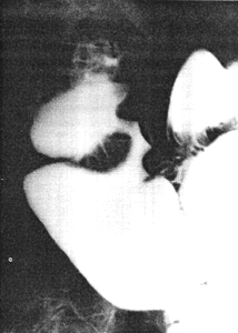
Physical examination revealed epigastric tenderness. A radiographic study, done
elsewhere, showed an irregular narrowing 3.5 cm in length at the pylorus (Fig.
31.1A); it had been interpreted as a carcinoma. Repeat radiological
examination showed that the narrowing was in reality a fistulous connection between the
distal end of the pyloric sphincteric cylinder and the base of the duodenal bulb on the
lesser curvature side. It was adjacent to, and located on the posterior aspect of the
pylorus. The sphincteric cylinder remained partially contracted as illustrated, neither
maximal contraction nor maximal expansion occurring. The duodenal bulb was
deformed. The condition was diagnosed as an acquired double pylorus, i.e. a
pyloroduodenal fistula as a result of a perforating ulcer. Endoscopy showed a benign
pyloric ulcer filled with necrotic material. It had perforated into the duodenum and the
instrument could be manipulated into the duodenum through the pylorus as well as
through the channel formed by the perforation.
A | B |
| Fig. 31.1 A,B.
Case S.K. A Narrow, irregular channel between pyloric sphincteric cylinder and
duodenal bulb, diagnosed as carcinoma. Normal pyloric aperture not filled with barium.
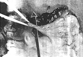
Cylinder partially contracted. B Resection specimen. Arrow through pyloroduodenal
fistula. Pyloric aperture visible behind arrow
|
At operation considerable fibrotic reaction was encountered and the duodenum had to be
dissected from the pancreas. A truncal vagotomy, antrectomy and Billroth I anastomosis
was performed. The resection specimen showed a second aperture between the stomach
and duodenum next to the normal pylorus, with a bridge of mucosal tissue between the 2
apertures (Fig. 31.1B) confirming the diagnosis of pyloroduodenal fistula or
so-called double pylorus.
Previous Page | Table of Contents | Next Page
© Copyright PLiG 1998


