

Go to chapter: 1 | 2 | 3 | 4 | 5 | 6 | 7 | 8 | 9 | 10 | 11 | 12 | 13 | 14 | 15 | 16 | 17 | 18 | 19 | 20 | 21 | 22 | 23 | 24 | 25 | 26 | 27 | 28 | 29 | 30 | 31 | 32 | 33 | 34 | 35 | 36 | 37 | 38 | 39
Chapter 3 (page 11)


Go to chapter: 1 | 2 | 3 | 4 | 5 | 6 | 7 | 8 | 9 | 10 | 11 | 12 | 13 | 14 | 15 | 16 | 17 | 18 | 19 | 20 | 21 | 22 | 23 | 24 | 25 | 26 | 27 | 28 | 29 | 30 | 31 | 32 | 33 | 34 | 35 | 36 | 37 | 38 | 39
Chapter 3 (page 11)
A B B |
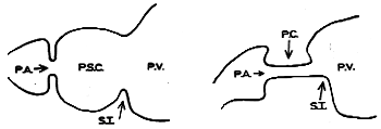
| Fig. 3.1. A. Divisions of pyloric region according to Cunningham. P.S.C., pyloric sphincteric cylinder; P.V., pyloric vestibule; P.A., pyloric aperture; S.I., sulcus intermedius. B Contracted pyloric sphincteric cylinder according to Cunningham. P.C., pyloric canal; P.V., pyloric vestibule; P.A., pyloric aperture; S.I., sulcus intermedius |
At the pyloro-duodenal junction the aboral margin of the muscular cylinder is increased
in thickness, thereby forming the massive muscular ring which encircles the pyloric
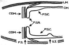
aperture. Cunningham (l906) called this ring the pyloric sphincteric ring (Fig. 3.2). The
ring protrudes into the commencement of the duodenum; when viewed from the
duodenal side, it presents as a smooth, rounded knob with a small puckered opening, the
pyloric aperture, in its centre.
The pyloric sphincteric ring is not a separate anatomical structure, but constitutes a
localized thickening of the cylinder, according to Cunningham (1906). On the gastric or
oral side, the circular fibres of the ring merge imperceptibly into those of the cylinder,
without any demonstrable anatomical boundary between the ring and cylinder. On the
aboral or duodenal side conditions are completely different. Here the circular fibres of
the ring are sharply demarcated from those of the duodenum by a fibrous septum; this
ensures a complete break between the circular musculature of the pylorus and that of the
duodenum (Fig. 3.2).
 | Fig. 3.2. Diagram of pyloric musculature according to Cunningham. P.S.C., pyloric sphincteric cylinder; P.S.R., pyloric sphincteric ring; F.S., fibrous septum; C.D.M., circular duodenal musculature; L.M., longitudinal musculature; C.M., circular gastric musculature; S.I., sulcus intermedius |
Not only the circular, but also the longitudinal fibres are present in greater mass in the
cylinder than in any other part of the stomach. In contrast to the circular fibres, a certain
percentage of gastric longitudinal fibres is continuous with those of the duodenum. The
more superficial fibres of the gastric longitudinal coat extend across the pyloro-duodenal
junction to merge with those of the duodenum. The deeper longitudinal fibres, as they
approach the pyloric aperture, dip into the sphincteric ring; some of these become
interwoven with the circular fibres of the ring, while others extend through the circular
coat to reach the submucosa.
On the oral side of the cylinder both its circular and longitudinal fibres merge
imperceptibly into those of the remainder of the gastric wall. Except for the palpable
thickening of the cylinder, and the shallow sulcus intermedius, no anatomical division
can be demonstrated on the oral side of the cylinder between its musculature and that of
the vestibule.
Cunningham (1906) called the muscular cylinder the pyloric sphincteric cylinder. The
aboral thickening, but integral part of the cylinder, was the pyloric sphincteric ring (Fig.
3.2). Contraction of the cylinder caused formation of the pyloric canal, which had to be
distinguished from the pyloric aperture.
He found minor variations in the arrangement of both circular and longitudinal muscle
fibres in different specimens. In one specimen for instance, all the longitudinal fibres
dipped into the sphincteric ring, while some superficial circular fibres of the ring were
carried on to the duodenum for a short distance. Sometimes the deeper longitudinal
fibres interlaced with the superficial circular fibres of the ring, forming a feltwork of
mixed fibres which was carried on to the duodenum. These variations soon gave way to
the proper coats of the duodenum, and in most specimens the arrangement was as
indicated above.
Previous Page | Table of Contents | Next Page
© Copyright PLiG 1998