


Go to chapter: 1 | 2 | 3 | 4 | 5 | 6 | 7 | 8 | 9 | 10 | 11 | 12 | 13 | 14 | 15 | 16 | 17 | 18 | 19 | 20 | 21 | 22 | 23 | 24 | 25 | 26 | 27 | 28 | 29 | 30 | 31 | 32 | 33 | 34 | 35 | 36 | 37 | 38 | 39
Chapter 23 (page 106)
Case 23.4. C.V., male aged 15 days, was admitted for projectile vomiting and
dehydration. Radiographic examination showed a constant narrowing, 1.5 cm in length,
in the pyloric region, with a concave indentation of the base of the duodenal bulb (Fig.
23.5). At times a small dilatation was seen in the centre of the narrowed canal. The
appearance is indicative of IHPS and can be explained as follows: the entire length of the
musculature of the pyloric sphincteric cylinder is hypertrophied, causing narrowing of the
lumen and a permanently contracted pyloric canal, as well as a concave indentation of the
base of the duodenal bulb. The dilatation in the centre of the narrowed canal is probably
an outward bulge between the right and left pyloric loops. The diagnosis was confirmed
at a Ramstedt operation a few days later.
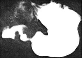 |
Fig. 23.5.
Case C.V. Permanently formed pyloric canal due to IHPS. Concave indentation base of bulb.
Small central dilatation in canal
|
Using a grey-scale unit with a 5MHz nonfocused transducer, Teele and Smith (l977) first
described the sonographic appearances in 5 babies with IHPS. By passing the transducer
transversely over the right lateral abdominal wall at the level of the costal margin, the
region of the pylorus was scanned through the liver; the hypertrophied pyloric
musculature presented as a round or oval echolucent mass with a central stellate
collection of echoes. The average antero-posterior diameter of each mass was 2.3 cm,
with a range of 1.8 to 2.8 cm. They were unable to identify a similar soft tissue mass in
normal babies. An echolucent mass could also be produced by the gastric "antrum" or
duodenal bulb filled with fluid, and by the hepatic flexure of the colon filled with stool,
but in those instances the appearance was evanescent and did not have the typical central
collection of echoes seen in IHPS.
Subsequently, various authors determined the measurements of the pyloric mass in IHPS
by direct viewing during ultrasonic examinations. Strauss et al. (l98l) examined 20
infants aged from 14 to 49 days with IHPS, initially using a static gray-scale B-scan and
later a real-time unit with a 5 MHz focused transducer. The pylorus was considered to be
abnormally thickened if it measured 1.5 cm or more in its ventral-dorsal diameter. (In
surgically controlled cases the pyloric mass of IHPS measured approximately 1.5 cm in
diameter). In 16 of the cases the mass measured from 1.5 cm to 3.0 cm. It presented as a
round anechoic mass with a central collection of echoes. In addition the pyloric "canal"
was seen to be elongated to 20 mm. Real-time scanning showed an absence of movement
of gastric contents across the pyloric canal.
Blumhagen and Coombs (l98l) examined 23 proven cases of IHPS between the ages of 2
and 10 weeks, using a B-scanner with a 6.0 mm focused 7.5 MHz transducer. In all cases
the hypoechoic single muscle layer was 4.0 mm thick or thicker. The thick hypoechoic
ring in the pyloric region was the sole criterion by which IHPS was diagnosed. The
fundamental advantage of ultrasonography over other methods of diagnosis was that it
visualized the hypertrophied muscle directly. Occasionally a hypoechoic ring might be
formed by the muscle layers of the normal "distal antrum" as seen on parasagittal sections
near the midline, but in those cases the layer was less than 4.0 mm in thickness. A false
positive sonograph might be obtained in pylorospasm, in which there was persistent
contraction of the circular musculature of the "distal antrum" and pylorus, creating a
cylindrical muscle mass. In those cases the muscle was also less than 4.0 mm in
thickness. The length of the hypertrophied muscle in IHPS was found to be variable,
with some patients having only a short segment of hypertrophy and others a more
elliptical mass.
Previous Page | Table of Contents | Next Page
© Copyright PLiG 1998