


Go to chapter: 1 | 2 | 3 | 4 | 5 | 6 | 7 | 8 | 9 | 10 | 11 | 12 | 13 | 14 | 15 | 16 | 17 | 18 | 19 | 20 | 21 | 22 | 23 | 24 | 25 | 26 | 27 | 28 | 29 | 30 | 31 | 32 | 33 | 34 | 35 | 36 | 37 | 38 | 39
Chapter 23 (page 105)

Go to chapter: 1 | 2 | 3 | 4 | 5 | 6 | 7 | 8 | 9 | 10 | 11 | 12 | 13 | 14 | 15 | 16 | 17 | 18 | 19 | 20 | 21 | 22 | 23 | 24 | 25 | 26 | 27 | 28 | 29 | 30 | 31 | 32 | 33 | 34 | 35 | 36 | 37 | 38 | 39
Chapter 23 (page 105)
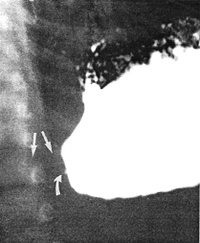 |
Fig. 23.2. Case B.G. Sharp cut-off pyloric region causing obstruction. Presumed extent of pyloric muscular hypertrophy (straight arrows). Central "beak" (curved arrow) |
Case 23.2. B.B., 28 day old male, had a history of post-feeds vomiting for the previous 2 weeks. Radiographic examination showed a severe, constant narrowing 2.0cm in length in the pyloric region of the stomach, causing obstruction. The narrowing was string-like in appearance and at its proximal end associated with concave indentations into the stomach (Fig. 23.3). The appearance is compatible with IHPS and can be explained as follows: hypertrophy of the musculature of the entire length of the pyloric sphincteric cylinder causes a pronounced narrowing of the lumen, which is permanent. Muscular hypertrophy surrounding the narrowed lumen indents the stomach, causing concave defects. The diagnosis of IHPS was confirmed at operation.
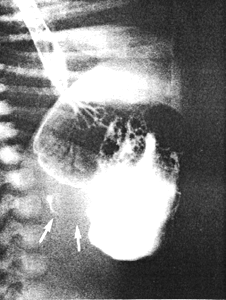 |
Fig. 23.3. Case B.B. String-like narrowing 2.0cm in length (arrows) due to pyloric muscular hypertrophy |
Case 23.3. M.M., male aged 10 days, was admitted with a history of vomiting since birth. Radiographic examination initially showed total obstruction of the pyloric region ("amputation of the antrum") (Fig. 23.4A). After 15 minutes, barium filled the pyloric canal, which was permanently contracted and approximately 2.0 cm in length (Fig. 23.4B); two longitudinal mucosal folds were seen in the contracted canal. There was a concave indentation of the base of the duodenal bulb. The appearance is that of IHPS and can be explained as follows: muscular hypertrophy of the entire length of the pyloric sphincteric cylinder initially obstructs the lumen; somewhat later filling of the permanently formed pyloric canal occurs. Muscular hypertrophy surrounding the latter indents the base of the duodenal bulb. Ramstedt operation two days later confirmed IHPS.
A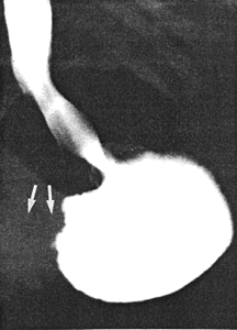 | B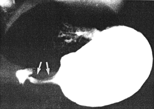 |
| Fig. 23.4. A Case M.M. Initial total obstruction pyloric region ("amputation of antrum"). Presumed extent of pyloric muscular hypertrophy (arrows). B Case M.M. Permanently formed pyloric canal, containing longitudinal mucosal folds, due to IHPS (arrows). Concave indentation base of duodenal bulb | |
Previous Page | Table of Contents | Next Page
© Copyright PLiG 1998