

Go to chapter: 1 | 2 | 3 | 4 | 5 | 6 | 7 | 8 | 9 | 10 | 11 | 12 | 13 | 14 | 15 | 16 | 17 | 18 | 19 | 20 | 21 | 22 | 23 | 24 | 25 | 26 | 27 | 28 | 29 | 30 | 31 | 32 | 33 | 34 | 35 | 36 | 37 | 38 | 39
Chapter 14 (page 69)


Go to chapter: 1 | 2 | 3 | 4 | 5 | 6 | 7 | 8 | 9 | 10 | 11 | 12 | 13 | 14 | 15 | 16 | 17 | 18 | 19 | 20 | 21 | 22 | 23 | 24 | 25 | 26 | 27 | 28 | 29 | 30 | 31 | 32 | 33 | 34 | 35 | 36 | 37 | 38 | 39
Chapter 14 (page 69)
 |
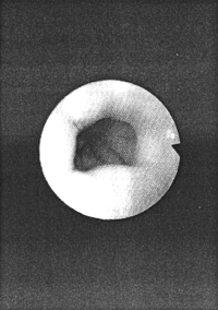
Fig. 14.1. Contraction ring formed by left pyloric loop. On caudal side of ring longitudinal mucosal folds recede into the distance, ending at pyloric aperture |
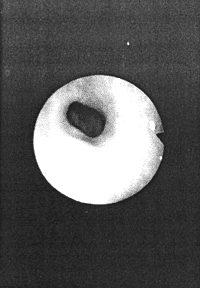
The region of the stomach between the ring (the left pyloric loop) and the pyloric aperture is the pyloric sphincteric cylinder. The ring may narrow (Fig.14.2) and contract to a narrow bore, through which mucus or at times mucosal folds may be retropelled. This indicates the stage of maximal contraction of the sphincteric cylinder as seen radiologically (Chap. 13).
 |
Fig. 14.2. Further narrowing of ring (left pyloric loop). The region beyond the ring and between it and the pyloric aperture (not visible) is the sphincteric cylinder |
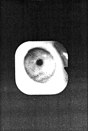
As the contraction causes the lumen to "disappear", the event itself (except for the contraction of the left loop) is not visible gastroscopically. In this sense maximal contraction of the pyloric sphincteric cylinder causes, gastroscopically speaking, an event horizon in the distal stomach. After a few seconds the sphincteric cylinder (including its left loop) relaxes, revealing the pyloric aperture, which is seen to be open (Fig. 14.3).
 |
Fig. 14.3. Pyloric aperture distal to the ring |
Should the tip of the gastroscope be closer to the pylorus, the pyloric aperture itself may
be seen to close or open to a certain extent, in the absence of contraction of the
sphincteric cylinder (marked air distension of the lumen may prevent contraction of the
cylinder). In this case the variation in size of the aperture is thought to be due to iris-like
action of the mucosal folds (Chap.13).
Previous Page | Table of Contents | Next Page
© Copyright PLiG 1998