

Go to chapter: 1 | 2 | 3 | 4 | 5 | 6 | 7 | 8 | 9 | 10 | 11 | 12 | 13 | 14 | 15 | 16 | 17 | 18 | 19 | 20 | 21 | 22 | 23 | 24 | 25 | 26 | 27 | 28 | 29 | 30 | 31 | 32 | 33 | 34 | 35 | 36 | 37 | 38 | 39
Chapter 13 (page 63)


Go to chapter: 1 | 2 | 3 | 4 | 5 | 6 | 7 | 8 | 9 | 10 | 11 | 12 | 13 | 14 | 15 | 16 | 17 | 18 | 19 | 20 | 21 | 22 | 23 | 24 | 25 | 26 | 27 | 28 | 29 | 30 | 31 | 32 | 33 | 34 | 35 | 36 | 37 | 38 | 39
Chapter 13 (page 63)
 |
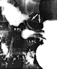
Fig. 13.17. Sphincteric cylinder contracted (arrows). The folds have changed in direction to become longitudinal. RPL, right pyloric loop; LPL, left pyloric loop. Note pseudodiverticulum between loops |
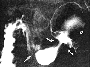
In some of our normal cases not only a change in direction, but also a cephalad
movement of the folds occurred during contraction of the sphincteric cylinder. The
following is an example:
 |
Fig. 13.18. Case G.O. Contraction of sphincteric cylinder and its right loop, closing the pyloric aperture (straight arrow). Retropulsion of mucosal folds (curved arrows) and barium (open arrow) |
Further evidence that the mucosa of the cylinder may move in an orad direction during its
contraction is seen in Chap. 36, cases 36.1 and 36.2. In these cases broad-based, sessile
mucosal polyps in the inactive, relaxed cylinder, moved orally during contraction of the
cylinder. The orad movement of the mucosa, while occurring normally, is not easily
demonstrable during the conventional barium studies, but retropulsion of barium during
contraction of the cylinder can often be shown; this should not be mistaken for
duodenogastric reflux (Chap. 27).
Further evidence of co-ordinated movements between the muscularis externa and mucosa
in the small bowel was presented by Sloan (l957). A correlation was found between the
direction of the mucosal folds on the one hand, and the degree of distension or
contraction of the walls on the other. During life longitudinal folds were seen to be
associated with peristaltic activity. This feature could not be reproduced in anatomical
specimens fixed in formalin.
Previous Page | Table of Contents | Next Page
© Copyright PLiG 1998