


Go to chapter: 1 | 2 | 3 | 4 | 5 | 6 | 7 | 8 | 9 | 10 | 11 | 12 | 13 | 14 | 15 | 16 | 17 | 18 | 19 | 20 | 21 | 22 | 23 | 24 | 25 | 26 | 27 | 28 | 29 | 30 | 31 | 32 | 33 | 34 | 35 | 36 | 37 | 38 | 39
Chapter 13 (page 62)
One of the functions of the stomach is to ensure transit and evacuation of chyme, which
is achieved by pressure gradients and motor movements. As indicated above, the
movements of the muscularis externa have been studied and analyzed in some detail.
The investigation of possible movements of the inner mucosal layer, on the other hand,
has been almost universally omitted in physiological and gastro-intestinal motility studies
(Chap. 2). This comes as a surprise, as the mucosa is in near contact with ingested solids,
liquids and the products of gastric digestion; only a layer of gastric mucus separates it
from the luminal contents. It seems reasonable to expect that the height and direction of
mucosal folds may have some bearing on the transit of contents, even if this were of a
purely mechanical nature.
For many years mucosal folds of the stomach were thought to be of little significance as
far as motility was concerned. The fundamental work of Forssell (l923, l939) has
received scant attention outside radiology, and it is an enigma of the medical literature
that little or no mention is made of these findings in other disciplines. Briefly, Forssell's
findings may be stated to be as follows:
Two independent but co-ordinated mechanisms of movement exist in the gastro-intestinal
tract, including the stomach, namely (1) movements of the muscularis externa, and (2)
movements of the mucosal coat, brought about by contractions of the muscularis
mucosae.
A certain contraction of the outer muscular tube is necessary for the formation of
macroscopic mucosal folds. The variability of the fold pattern is also dependent on the
hydrodynamic action of the fluid content of the submucosa, which in turn depends on the
degree of filling of the blood vessels; the mass of mucous membrane, and consequently
the volume of its folds, is regulated by the varying vascularity in the submucosa.
In addition Forssell showed that the surface of the mucosa, or mucosal relief pattern, may
vary from moment to moment; these movements are independent of, but co-ordinated
with the contractions of the muscularis externa. Especially in the small intestine, but also
in the stomach, various active contractile shapes, in some of which the mucosa "grips"
particles of food, may be discerned. These consist of digestion chambers, blocking or
filtering devices, re-absorption reservoirs and smooth or corrugated transporting tubes.
In this way each region may best meet the varying demands placed on it from moment to
moment, namely digestion, storage, absorption and transport respectively. One moment
the mucosa may be occupied with one task, the next with another. Forsell called the
inherent ability of the mucosa to move "mucosal autoplastik", providing a working relief
pattern. It also determined to a large extent the number, position and form of the folds.
While the coarser breakdown of food particles is effected by contractions of the
muscularis externa, the finer dispersion occurs through changes in the relief pattern of the
mucosa, which may enhance or counteract effects of contraction of the muscular walls.
The special contractile organ of the mucous membrane is the muscularis mucosae; being
attached to the mucosa and being incorporated in the submucosa, it is able to displace the
former in different directions.
While elaborating on Forssell's work, Golden (l937), during radiological examinations,
observed that in some cases the mucosal folds in the "antrum" ran irregularly transverse
to the long axis, but when "antral systole" occurred they changed in direction and came to
lie parallel to the long axis. It was surmized that during this process the mucosa also
moved in an oral direction; otherwise, if this failed to occur, the folds would be
exaggerated and jammed toward the pylorus during contraction of the canalis, tending to
cause obstruction. The change in direction of folds only occurred during complete
contractions of the canalis which expelled gastric contents.
Brooks et al. (l948), while investigating the gastric mucosa in canines, confirmed
Forssell's view that the gastric mucosa was capable of independent movements.
However, contraction of the muscularis externa also produced changes in the mucosal
pattern. No clear picture of co-ordinated movements between the muscularis externa and
muscularis mucosae emerged from their study.
Evidence of co-ordinated movements in the small bowel was furnished by Deucher
(l951), who noted at operations that mucosal folds were transverse to the long axis in
distended regions, and longitudinal in contracted segments of the bowel.
In radiological and experimental anatomical studies of mucosal fold movements Keet
(l974, l978) found that normally, transverse or oblique mucosal folds could be
demonstrated in the pyloric sphincteric cylinder while the latter was distended or relaxed
(Fig. 13.16). (Comment: The terms "transverse" and "oblique" indicate the
direction in relation to the long axis. Folds which appear transverse on the two-
dimensional radiographic image are in reality circular, surrounding the tube-like lumen.
Oblique folds are of a spiral nature).
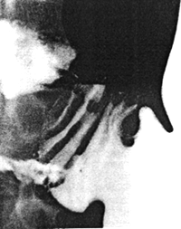 |
Fig. 13.16.
Pyloric sphincteric cylinder partially distended. All its mucosal folds are
transverse (i.e. circular)
|
Previous Page | Table of Contents | Next Page
© Copyright PLiG 1998