

Go to chapter: 1 | 2 | 3 | 4 | 5 | 6 | 7 | 8 | 9 | 10 | 11 | 12 | 13 | 14 | 15 | 16 | 17 | 18 | 19 | 20 | 21 | 22 | 23 | 24 | 25 | 26 | 27 | 28 | 29 | 30 | 31 | 32 | 33 | 34 | 35 | 36 | 37 | 38 | 39
Chapter 13 (page 58)
Finally, a simultaneous widening of the two legs or loops of the inverted V occurs,
causing the disappearance of the pseudodiverticulum. This results in a triangular region
of tight contraction (Fig. 13.10). The pyloric canal is now fully formed and runs through
the contracted region as a thin channel containing one or more barium-lined longitudinal
mucosal furrows.
 |
Fig. 13.10.
Sketch of maximal contraction. D.B., duodenal bulb; P.C., pyloric canal; M.C.,
muscular contraction
|
The events on the lesser and greater curvatures, together with the narrowing of the lumen,
occur simultaneously in one smooth, integrated, uninterrupted movement.
This then constitutes a maximal contraction of the distal 2.0 to 3.0 cm of the stomach. It
occurred in all normal cases. After two to three seconds the contraction relaxed, the
lumen reassumed its "resting" diameter, and the process was repeated, showing it to be of
cyclical nature. A "pyloric cycle" denotes the time from commencement of one
contraction to the commencement of the next.
The frequency of pyloric cycles per minute was determined as follows: In 50 of the
subjects a stage was awaited in which maximal contractions occurred regularly. At the
commencement of one of these contractions an assistant with a stopwatch would be told
to "start!". At the end of 30 seconds the assistant would exclaim "stop!". The number of
cyclical contractions per 30 second period per subject would be counted, from which the
average number of contractions per minute per subject could be established. This proved
to be approximately three and a half cycles of contraction per minute. (Because of
various factors, e.g. the delay in responding to "start" and "stop", the correct figure is
estimated to be somewhat less and probably between 3 and 3Æ cyles per minute).
A maximal contraction wave was associated with a sharp increase in intraluminal
pressure, ranging up to 34 mm Hg (vide supra: See also Chapter 15).
An attempt was made to correlate details of the contractions of the distal 3.0 to 4.0 cm of
the stomach, as revealed by radiology, with the muscular anatomy as described by
Cunningham (l906), Forsell (l913), Cole (l928) and Torgersen (l942). The findings,
which were analyzed in 320 cases (Keet l957), can be described as follows:
From the validation studies it is concluded that dynamic narrowing of the barium-
containing lumen, as seen during a maximal contraction, is caused by muscular
contraction of the walls. In order to visualize the shape and extent of muscular
contraction, the method of focussing on the "black" areas of contraction surrounding the
"white" barium-filled lumen is used. Using this perspective, it appears that the arrival of
a peristaltic wave at a point 3.0 to 4.0 proximal to the pyloric aperture, initiates
contraction of the various divisions of the pyloric sphincteric cylinder; normally this
progresses uninterruptedly to culminate in a tight, maximal contraction of the entire
cylinder.
One of the first events to occur, namely widening of the pyloric ring on the lesser
curvature side (Fig. 13.8), tallies with commencing contraction of the pyloric muscle
torus or knot, which is located in this situation. (This is also evident on the radiograph
shown in Fig. 13.6).
Commencing formation of a gastric loculus on the greater curvature side (Fig. 13.8)
tallies with early contraction and approximation of the right and left pyloric loops (the
latter is adjacent to the stationary peristaltic wave on the greater curvature).
On the lesser curvature the indentation caused by continuing contraction of the muscle
torus fuses with that of the stationary peristaltic wave, to cause a single region of
contraction (Fig. 13.9). At this stage the right and left pyloric loops radiate in a fan-like
shape from the muscle torus to surround the greater curvature, where two contraction
rings are seen. The rings compress the gastric loculus, resulting in the formation of a
physiological pseudodiverticulum (Fig. 13.9) (see also Fig. 13.11).
 |
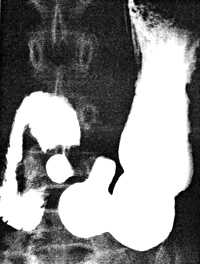
Fig. 13.11.
Radiograph of normal, physiological pseudodiverticulum. Note single area of
contraction on lesser curvature and two loops on greater curvature
|
Continuing contraction of the muscle torus and the two loops further compresses the
pseudodiverticulum, causing its disappearance and resulting in a single, cylindrical region
of tight contraction (Fig. 13.10). The compressed lumen at this stage is not more than
2-3 mm in diameter; it extends through the centre of the maximally contracted
cylinder as a thin tube, often containing one or more longitudinal mucosal furrows (see
Fig. 13.15B).
It is probable that narrowing of the lumen is brought about by contraction of the circular,
and approximation of the loops by contraction of the longitudinal muscle fibres of the
sphincteric cylinder.
Previous Page | Table of Contents | Next Page
© Copyright PLiG 1998


