


Go to chapter: 1 | 2 | 3 | 4 | 5 | 6 | 7 | 8 | 9 | 10 | 11 | 12 | 13 | 14 | 15 | 16 | 17 | 18 | 19 | 20 | 21 | 22 | 23 | 24 | 25 | 26 | 27 | 28 | 29 | 30 | 31 | 32 | 33 | 34 | 35 | 36 | 37 | 38 | 39
Chapter 13 (page 57)
A caudally travelling peristaltic wave was never seen to proceed as far as the pyloric
aperture.
The degree (or range) of contraction (which would probably correspond to amplitude in
manometry) of the distal 3.0 to 4.0 cm long cylinder varied. During a single examination
various degrees of contraction might be seen, e.g. feeble or incomplete contractions
might occur in the early stages of the examination (Fig. 13.7), followed by maximal or
complete contractions in which the lumen was bisected, later on (see also Chapter 12).
During an examination contractions might become shallower for various (and sometimes
unknown) reasons. For instance, not uncommonly contractions became very shallow, or
even disappeared temporarily, after the patient had been supine and re-assumed the erect
position. As a rule, however, once maximal contractions started, they occurred fairly
regularly.
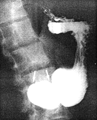 |
Fig. 13.7.
Incomplete cylindrical (or "systolic") contraction (arrows) of distal 3-4 cm of stomach.
|
Each maximal (or complete) cylindrical contraction of the distal 3.0 to 4.0 cm of the
stomach was initiated by a peristaltic wave.
In all normal cases the contractions conformed to a definite pattern. The details may best
be visualized if attention is focussed on the "black" regions of contraction surrounding
the barium filled lumen. With this purpose in mind, sketches of radiographs taken at
various stages have been made. The details of a typical maximal contraction may be
described as follows:
When the peristaltic wave comes to a halt 3.0 to 4.0 cm orally to the pyloric ring, a
widening of the indentation of the ring on the lesser curvature side occurs (Fig. 13.8). On
the greater curvature a loculus of gastric lumen begins to form between the ring and the
indentation of the arrested peristaltic wave (Fig. 13.8). While the peristaltic wave
remains stationary, its indentation widens progressively. Simultaneously the indentation
of the ring on the greater curvature also widens. The widening of these indentations can
only be due to muscular contraction, and the effect is to compress the loculus so that it
becomes smaller and resembles what can be called a pseudodiverticulum (Fig. 13.9).
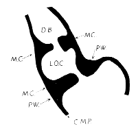 |
Fig. 13.8.
Sketch of commencing normal contraction showing widening of pyloric ring
on lesser curvature side. D.B., duodenal bulb; L.O.C., loculus of gastric lumen; M.C.,
muscular contraction; P.W., stationary peristaltic wave; C.M.P., circular muscularis
propria
|
Meanwhile the indentation on the lesser curvature enlarges further and merges
imperceptibly with the impression of the original peristaltic wave which remained
stationary at this point (Fig. 13.9). The effect of this is to cause a single, wide region of
contraction on the lesser curvature, as opposed to two separate contractions on the greater
curvature. The contracted region at this stage resembles an inverted V, with the apex on
the lesser curvature and the two limbs radiating to and surrounding the greater curvature.
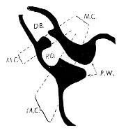 |
Fig. 13.9.
Sketch of continuing normal contraction. D.B., duodenal bulb; P.D., pseudodiverticulum;
M.C., muscular contraction; P.W.; stationary peristaltic wave
|
Previous Page | Table of Contents | Next Page
© Copyright PLiG 1998