

Go to chapter: 1 | 2 | 3 | 4 | 5 | 6 | 7 | 8 | 9 | 10 | 11 | 12 | 13 | 14 | 15 | 16 | 17 | 18 | 19 | 20 | 21 | 22 | 23 | 24 | 25 | 26 | 27 | 28 | 29 | 30 | 31 | 32 | 33 | 34 | 35 | 36 | 37 | 38 | 39
Chapter 13 (page 53)
Narrow, circumferential indentations of the barium column appeared in the body of the
stomach and proceeded to move in a caudal direction. Opposite the indentations the fine,
flexible wires remained in their original position, showing that these indentations were
due to contraction waves in the walls and not "falling together" of the walls. At a point
3.0 to 4.0 cm orally to the pyloric ring each wave became stationary, at the same time
initiating a concentric, cylindrical narrowing of the barium column in the remaining part
of the stomach, as far as and including the area of the ring. Again the wires were seen to
remain in their original position. During a contraction the space between the wires and
the luminal barium widened to approximately 8.0 to 10.0 mm all round, indicating an
active, tube-like or cylindrical contraction of the muscular walls, 3.0 to 4.0 cm in length
(Fig. 13.3). After a second or two of maximal contraction, the walls relaxed and the
cycle was repeated.
On completion of the radiological examination the wires were removed by gentle traction
on their external ends. None of the patients suffered any discomfort or untoward
sequelae; recovery was normal.
It is concluded that the narrowing of the intraluminal barium column was not due to a
passive falling together of the walls, as the fine, flexible wires on the serosal surface
remained in their original positions. The narrowing of the column was due to active
contraction of the walls between the serosa and the barium containing lumen.
(Endoscopic ultrasonography confirms that "peristaltic" contraction of the wall produces
wall thickening, as mentioned in Chapter 10).
According to Code and Carlson (l968) the stomach has 3 functional regions
corresponding to its anatomic divisions, namely the fundus, corpus and pyloric antrum,
which, in their view, is the region stretching from the incisura angularis to the pylorus.
(Comment: While the concept of these "anatomical" divisions appears to be
widely accepted, it is shown in Chapters 2 and 3 that the division is of an arbitrary nature
and not based on anatomical facts). In terms of motor activity Code and Carlson (l968)
divided the antrum into two segments of varying length. The caudal portion participates
in a simultaneous segmental contraction called the terminal antral contraction (TAC),
previously described by Carlson, Code and Nelson (l966). The cephalad portion of the
antrum, according to these authors, is not usually involved in TAC; however, at times
TAC involves only the distal one to two centimeters of the antrum, and at other times
almost the entire antrum. During TAC a simultaneous contraction of the terminal
segment of the antrum occurs, a phenomenon which corresponds to antral systole
previously described by Golden (l937). The cephalad and terminal (or caudal) segments
of the antrum, together with the pylorus (also called the pyloric canal or pyloric
sphincter) constitute a functional motor unit according to Code and Carlson (1968); the
separate parts vary in their dimensions but contract in a co-ordinated way. The pyloric
canal closes vigorously with contraction of the terminal antrum. (Comment:
According to these authors the pyloric ring constitutes the pyloric sphincter. The pyloric
canal is equated with the aperture).
Ruch and Patton (l973) state that morphologically, histologically and functionally the
stomach is divided into 3 parts, viz. the fundus, corpus and pyloric antrum or pars
pylorus, a narrower, more muscular, non-acid secreting region. The fundus and corpus
together form a somewhat bulbous, thin-walled storage and secretory chamber, while
food is fragmented and mixed with digestive juices in the "antrum". There is no
structural discontinuity between these regions, which are only modifications of a basic
pattern.
From the point of view of motility, other authors divided the stomach into two parts,
namely a proximal one third and a distal two thirds (Kelly l98l; Funch-Jensen l987). The
division was said to be based not on the usually accepted anatomical considerations, but
on the type of smooth muscle activity. During swallowing the proximal part of the
stomach relaxes, which allows filling without a significant increase in pressure. It acts as
a receptacle and determines to a large extent the emptying of liquids. The distal two
thirds shows active peristalsis which propagate luminal contents towards the pylorus,
thereby effecting the emptying of solids.
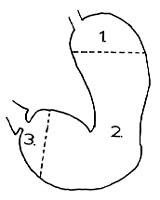
In terms of motor function, based on the muscular anatomy, radiologically visible
contraction patterns, manometrically recordable pressure waves and myoelectric activity,
it is our view that the stomach should be divided into three parts namely (1) the fornix,
(2) the corpus and sinus and (3) the distal 3.0 to 4.0 cm (Keet l957) (Fig. 13.4).
 |
Fig. 13.4.
In terms of motor activity the stomach should be divided into three parts. 1.,
fornix; 2., corpus and sinus; 3., distal 3-4 cm
|
Previous Page | Table of Contents | Next Page
© Copyright PLiG 1998


