

Go to chapter: 1 | 2 | 3 | 4 | 5 | 6 | 7 | 8 | 9 | 10 | 11 | 12 | 13 | 14 | 15 | 16 | 17 | 18 | 19 | 20 | 21 | 22 | 23 | 24 | 25 | 26 | 27 | 28 | 29 | 30 | 31 | 32 | 33 | 34 | 35 | 36 | 37 | 38 | 39
Chapter 13 (page 52)
In the second and third parts of the duodenum the base line again represented
intraluminal pressure in the absence of radiologically visible motor activity. Two waves
of pressure increase were noted manometrically:
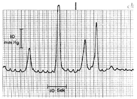
- Nonrhythmic, simple, brief monophasic waves, causing intraluminal pressure
increases of 4.0 to 35 mm Hg (the majority being in the 7.0 to 34 mm Hg range),
and lasting 2 to 8 seconds (Fig. 13.2). These waves occurred repeatedly in all
subjects and conform to Type I duodenal waves (Foulk et al. l954; Vantrappen et
al. l965; Friedman et al l965); it was suggested that they should be designated
phasic waves (Texter l968).
- In two of the subjects nonrhythmic, complex waves consisting of a rise in base
line pressure of 3.0 to 4.0 mm Hg and lasting from 40 seconds to 2½
minutes, with superadded peaks of l9 to 23 mm Hg lasting for 5 to 6 seconds,
were seen occasionally. These conform to Type III duodenal waves (Foulk et al.
l954; Vantrappen et al. l965; Friedman et al. l965); it was suggested that they
should be designated tonic waves (Texter l968).
During both types of waves radiologically visible, circumferential narrowing of
the luminal barium column occurred simultaneously with the increases in pressure
(see also Chap. 15).
 |
Fig. 13.2.
Four monophasic duodenal pressure waves. Each was associated with a
radiologically visible contraction. During each wave mucosal folds changed in direction
to become longitudinal. Base line indicates intraluminal pressure in absence of motor
activity. Ten-second marker on zero line
|
It was concluded that the narrowing of the intraluminal barium column was due to active
contraction of the walls.
The living anatomy was investigated in a number of patients who had to undergo
cholecystectomy during the ordinary course of events (Keet and Heydenrych l982). The
investigation was designed to determine the spatial relationship between the barium
column in the lumen and the walls.
Ethical Considerations.
The study was undertaken in informed, adult, white, volunteer patients who had been
admitted to hospital with definite indications for cholecystectomy. All aspects of the
procedure had been considered carefully beforehand by ourselves, our peers and the Head
of the Department of Surgery; no objections were raised. The Ethical Committee
indicated that it could find no objection to the procedure.
Six patients were examined. On completion of the cholecystectomy, and before closure
of the abdomen, the stomach and duodenum were shown to be normal by means of direct
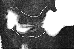
inspection and palpation. Two fine, flexible stainless metal wires, similar to the wires
used in the leads of myocardial pacemakers, were attached to the serosal surface of the
pyloric region of the stomach and first part of the duodenum by means of superficial,
interrupted, absorbable sutures (Fig. 13.3). One wire was attached to the lesser and the
other to the greater curvature, the free "duodenal" ends of both wires being brought to the
surface (as in the case of a postoperative T-tube) through the cholecystectomy incision,
which was subsequently closed in the usual way.
Approximately 8 days later, on the day before discharge, each patient had a limited
radiographic study as follows: after an overnight fast 4 to 5 mouthfulls of the usual liquid
barium suspension was swallowed in the erect position, so as to outline the horizontal
part of the gastric lumen and to extend well up into the vertical part. The space between
the metal wires on the serosal surfaces and the luminal barium indicated the thickness of
the wall; during the motor quiescent stage it was approximately 4.0 to 5.0 mm. After
emptying into the duodenum had commenced, gastric contractions were studied by means
of radiographic TV monitoring and appropriate radiographs.
 |
Fig. 13.3.
Radiograph showing living anatomy. Two fine, flexible metal wires
(retouched) are attached to serosal surfaces of lesser and greater curvatures. The space
between the wires and intraluminal barium indicates the cylindrical muscular contraction
|
Previous Page | Table of Contents | Next Page
© Copyright PLiG 1998


