The Pyloric Sphincteric Cylinder in Health and Disease


Go to chapter: 1 | 2 | 3 | 4 | 5 | 6 | 7 | 8 | 9 | 10 | 11 | 12 | 13 | 14 | 15 | 16 | 17 | 18 | 19 | 20 | 21 | 22 | 23 | 24 | 25 | 26 | 27 | 28 | 29 | 30 | 31 | 32 | 33 | 34 | 35 | 36 | 37 | 38 | 39
Chapter 5 (page 22)


Border on Aboral Side

Using macroscopic techniques in 53 fresh, non-diseased post-mortem specimens, Landboe-Christensen (l944) found that the border between pyloric and duodenal mucosa might lie either on the perpendicular, duodenal side of the pyloric ring, at the top of the ring, or on the sloping gastric side of the ring. With ageing there was a tendency for the border to shift from the duodenal to the gastric side of the ring.

Using microscopic methods in 110 cases of partial gastrectomy for duodenal ulcer, Oi and Sakurai (l959) came to essentially similar conclusions. In all specimens the border was within 5.0 mm of the summit of the pyloric "sphincter muscle" (presumably the pyloric ring), either on the duodenal or on the gastric side. The pyloric mucosa was in direct contact with the duodenal mucosa, i.e. the border could be identified as a line, in contrast to the border between pyloric and oxyntic mucosa, which consisted of a transitional zone of varying width.

Border on Oral Side

Landboe-Christensen (l944) studied the extent of the pyloric mucosal zone on the oral side by macroscopic tehcniques in 47 non-diseased, fresh post-mortem specimens. In all instances this zone was longer on the lesser than on the greater curvature, covering on average 7.2 cm of the lesser, and 5.2 cm of the greater curvature. The average percentage of the lesser curvature covered was 44 percent, and the average percentage of the greater curvature covered was 12 percent. The border between pyloric and oxyntic mucosa was irregular, often dentate or tortuous, and as a rule a number of islets of oxyntic mucosa were encountered in the pyloric zone. There was a transitional area of varying width between the two zones. In practice, a line drawn from the junction of the upper three- fifths and lower two-fifths of the lesser curvature, running downwards and to the right, indicates the approximate boundary of pyloric mucosa (Fig. 5.1).

Dean and Mason (l964) studied 117 fresh gastric resection specimens microscopically (resected for either duodenal or gastric ulcer). In l8 cases of duodenal ulcer the average length of pyloric mucosa along the lesser curvature was 9.0cm, and along the greater curvature 7.4 cm. On an average the pyloric mucosa extended for l.5cm further up the lesser than the greater curvature. In the majority of all duodenal ulcer specimens the transitional zone between pyloric and oxyntic mucosa varied from 0.5 to 5.0 cm in width, the average width being 1.0cm. The upper limit of the pyloric mucosa showed wide individual variations. Patients with gastric ulcers tended to have a greater extent of pyloric mucosa than those with duodenal ulcers, and women with gastric ulcers had more pyloric mucosa than men with gastric ulcers.

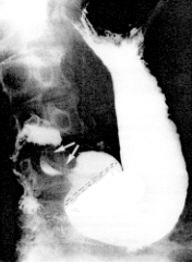
Fig. 5.1. Relationship between pyloric sphincteric cylinder and pyloric mucosal zone in normal stomach. Arrows, contracted sphincteric cylinder; broken line, approximate border between pyloric and oxyntic mucosal zones.

Schrager et al. (l967) studied the "antrum" microscopically in 45 normal stomachs obtained at necropsy, in 75 resection specimens of duodenal ulcer and in 40 specimens of gastric ulcer. Normal stomachs showing clear histology were not easily obtained due to the speed with which autolysis occurs after death; in only 13 could measurements be made. Normally the average length of pyloric mucosa between the "sphincter" (i.e. the pyloric ring) and the boundary zone on the lesser curvature was 8.9 cm, with an average "sphincter" to cardia distance of 22.5 cm. Thus the pyloric zone encompassed 40 percent of the lesser curvature. On the greater curvature the average figures were 4.8 cm, 39 cm and 12 percent respectively. Comparative studies showed that in the majority of duodenal ulcer patients the pyloric zone was larger than in normal controls, and in the majority of gastric ulcer cases it was almost twice the normal size.

In surgical pH monitoring tests, Capper et al. (l962, l966) found a wide variation in size between the small pyloric zone of duodenal ulcer and the larger zone of gastric ulcer cases. With duodenal ulceration the zone was usually of normal size or smaller, extending to a line 3.0 to 4.0 cm from the pylorus. (In their only normal subject, the pyloric zone was 4.0 to 5.0 cm in length.) In gastric ulcer cases the pyloric zone was very much larger, at times encompassing the whole of the lesser curvature. Capper et al. concluded that the junction between the distal alkaline, pyloric zone and the proximal oxyntic zone was not static, but that it might migrate up and down the stomach. The alkaline zone could have been a normal "antrum", but it could also have represented oxyntic mucosa which had been altered by gastritis.


Previous Page | Table of Contents | Next Page
© Copyright PLiG 1998