Go to chapter: 1 | 2 | 3 | 4 | 5 | 6 | 7 | 8 | 9 | 10 | 11 | 12 | 13 | 14 | 15 | 16 | 17 | 18 | 19 | 20 | 21 | 22 | 23 | 24 | 25 | 26 | 27 | 28 | 29 | 30 | 31 | 32 | 33 | 34 | 35 | 36 | 37 | 38 | 39
Chapter 38 (page 191)
In three of our verified cases the size of the defect in the duodenal bulb was seen to be
related to the degree of contraction of the pyloric sphincteric cylinder (Keet 1952). In
these cases a shallow defect might be visible during relaxation or inactivity of the
cylinder. During contraction the duodenal defect became more extensive, reaching its
greatest volume with maximal contraction of the cylinder. The same feature was seen in
most of our unverified cases as well. It was not unusual for the duodenal bulb to appear
normal prior to contraction of the cylinder, with the defect only appearing during
contraction.
In contrast, Stiennon (1960) noted that tortuous prepyloric rugae sometimes "prolapsed
backward", i.e. in an orad direction, through an advancing peristaltic wave. In most cases
a somewhat stronger or tighter wave finally forced the mucosa through the pylorus. A
similar phenomenon was described by White et al. (1966), who reported that the
hyperaemic and oedematous prepyloric folds pushed back into the stomach as the
"sphincter" closed. It was shown in Chapter 13 that orad movement of the mucosa in the
pyloric sphincteric cylinder may occur normally during contraction of the cylinder.
Furthermore sessile mucosal polyps in the cylinder may move in an orad direction during
contraction of the cylinder (Chap. 36).
Levin (l97l) again stated that the duodenal defect of prolapsed gastric mucosa became
more prominent during "gastric systole", and that the shape of the bulb reverted to normal
during "gastric diastole".
The following are examples of further cases encountered by us:
Case Reports
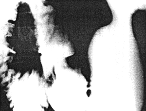
Case 38.1 J.S., 35 year old male had intermittent, burning epigastric pain apparently
related to meals, for the previous 5 years. Large meals occasionally caused vomiting.
Clinical examination was negative. Radiology revealed no abnormality in the
oesophagus and stomach. During phases of distension or partial contraction of the
pyloric sphincteric cylinder, the duodenal bulb appeared normal (Fig.
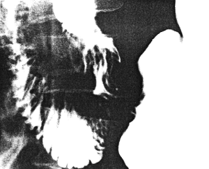
38.1A). With maximal contraction of the sphincteric cylinder an umbrella-
like or mushroom-like defect appeared in the base of the duodenal bulb; it was
continuous with longitudinal mucosal folds extending through the fully formed pyloric
canal (Fig. 38.1B), and the diagnosis of prolapse of gastric mucosa into the
duodenum was made. Gastroscopy showed a few prominent mucosal folds which
appeared to be redundant and capable of prolapsing into the duodenum. No evidence of
ulceration or other pathology was seen.
|
Fig. 38.1 A.
Case J.S. Partial contraction of pyloric sphincteric cylinder.
Base of duodenal bulb normal
|
|
Fig. 38.1 B.
Case J.S. Maximal contraction of sphincteric cylinder.
Umbrella-like defect base of bulb, continuous with longitudinal mucosal folds in pyloric
canal
|
Case 38.2 A.A., 20 year old male, a known case of duodenal ulceration, had received
anti-ulcer therapy for the preceding year. Because of a recurrence of symptoms
radiographic examination was requested.
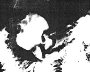
Several prominent, tortuous mucosal folds were seen in the pyloric sphincteric cylinder
while it was relaxed; the base of the duodenal bulb appeared normal, but there was a
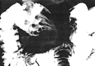
possible ulcer near its apex (Fig. 38.2A). During maximal contraction of the
pyloric sphincteric cylinder an umbrella-like defect appeared in the base of the duodenal
bulb, continuous with longitudinal gastric mucosal folds stretching through the fully
formed pyloric canal (Fig. 38.2B). The case was diagnosed as prolapse of
gastric mucosa and probable active duodenal ulceration. Control double contrast
radiographic examination a month later failed to show the prolapse. At this examination
administration of an anticholinergic substance relaxed the gastric walls and insufflation
of air caused luminal distension, factors which prevented the sphincteric cylinder from
contracting.
|
Fig. 38.2 A.
Case A.A. Prominent, tortuous mucosal folds in relaxed pyloric
sphincteric cylinder. Base of duodenal bulb normal. Possible duodenal ulcer
|
|
Fig. 38.2 B.
Case A.A. Maximal contraction of sphincteric cylinder. Umbrella-like defect base of bulb,
continuous with longitudinal mucosal folds in pyloric canal
|
Previous Page | Table of Contents | Next Page
© Copyright PLiG 1998