


Go to chapter: 1 | 2 | 3 | 4 | 5 | 6 | 7 | 8 | 9 | 10 | 11 | 12 | 13 | 14 | 15 | 16 | 17 | 18 | 19 | 20 | 21 | 22 | 23 | 24 | 25 | 26 | 27 | 28 | 29 | 30 | 31 | 32 | 33 | 34 | 35 | 36 | 37 | 38 | 39
Chapter 37 (page 186)


Go to chapter: 1 | 2 | 3 | 4 | 5 | 6 | 7 | 8 | 9 | 10 | 11 | 12 | 13 | 14 | 15 | 16 | 17 | 18 | 19 | 20 | 21 | 22 | 23 | 24 | 25 | 26 | 27 | 28 | 29 | 30 | 31 | 32 | 33 | 34 | 35 | 36 | 37 | 38 | 39
Chapter 37 (page 186)
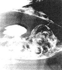 |
Fig. 37.2. Case T.M. Absent cyclical activity of pyloric sphincteric cylinder. Pyloric aperture patulous. Retention of food residues and 8.0mm diameter tablets |
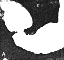
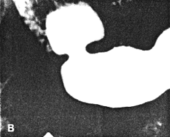
Case 37.3. F.J., 64 year old female with longstanding, non-insulin dependent diabetes mellitus had received oral anti-diabetic therapy for 8 years. She was admitted for dyspepsia, epigastric pain and diabetic retinopathy. Upper gastrointestinal radiological examination showed a small benign-looking gastric polyp immediately below the gastro- oesophageal junction. Gastric peristaltic waves were shallow and appeared sluggish. The pyloric sphincteric cylinder was partially contracted throughout the examination; while minor degress of movement of the walls were discernable, there was a total lack of cyclical contraction and relaxation, the appearance remaining more or less unchanged (Fig. 37.3). The pyloric aperture remained patent, having a diameter of approximately 1.0 cm; continuous emptying of fluid barium occurred through the patulous pylorus. At gastroscopy a small, benign polyp was removed. The remainder of the stomach, the pylorus and duodenum showed no lesion. The motility disturbance of the pyloric sphincteric cylinder with the patulous pyloric aperture was thought to be compatible with diabetic gastroparesis.
 |  |
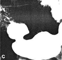 | 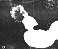 |
| Fig. 37.3. A-D Case F.J. Pyloric sphincteric cylinder permanently contracted. Cyclical activity absent. Pyloric aperture patulous with diameter of 1.0 cm | |
Case 37.4. M.J., 63 year old female was admitted for ischaemic heart disease, loss of weight and anorexia. She was a known case of non-insulin dependent diabetes mellitus and had received oral therapy for the previous 10 years. There was severe target organ involvement with diabetic retinopathy. Upper gastro-intestinal radiological examination showed a constant contraction of the pyloric sphincteric cylinder which remained unchanged throughout the examination (Fig. 37.4); this was thought to be compatible with early diabetic gastroparesis.
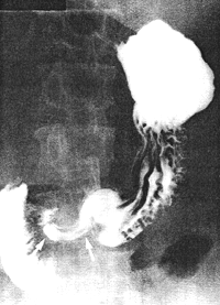 |
Fig. 37.4. Case M.J. Permanent contraction of pyloric sphincteric cylinder (arrows) |
Previous Page | Table of Contents | Next Page
© Copyright PLiG 1998