

Go to chapter: 1 | 2 | 3 | 4 | 5 | 6 | 7 | 8 | 9 | 10 | 11 | 12 | 13 | 14 | 15 | 16 | 17 | 18 | 19 | 20 | 21 | 22 | 23 | 24 | 25 | 26 | 27 | 28 | 29 | 30 | 31 | 32 | 33 | 34 | 35 | 36 | 37 | 38 | 39
Chapter 35 (page 178)


Go to chapter: 1 | 2 | 3 | 4 | 5 | 6 | 7 | 8 | 9 | 10 | 11 | 12 | 13 | 14 | 15 | 16 | 17 | 18 | 19 | 20 | 21 | 22 | 23 | 24 | 25 | 26 | 27 | 28 | 29 | 30 | 31 | 32 | 33 | 34 | 35 | 36 | 37 | 38 | 39
Chapter 35 (page 178)
 |
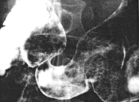
Fig. 35.2. Case M.B. Double contrast examination. Constant contraction of pyloric sphincteric cylinder with absent cyclical activity. Pyloric aperture patent. |
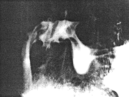
Case 35.3 M.A., female aged 50 years, presented with dysphagia of one year's
duration. Radiographic examination showed a carcinoma 5.0 cm in length in the lower
oesophagus. There was marked contraction or spasm of the entire pyloric sphincteric
cylinder, with a prominent pseudo-diverticulum on its greater curvature side (Fig. 35.3).
Occasionally a minor degree of movement was seen; most of the time the appearance
remained as indicated. Subsequent oesophagoscopies confirmed carcinomatous
involvement of the lower third of the oesophagus.
 |
Fig. 35.3. Case M.A. Constant, near maximal contraction of pyloric sphincteric cylinder with pseudo-diverticulum on greater curvature side. |
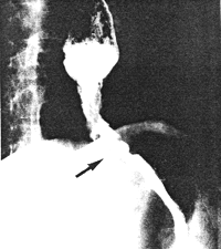
Case 35.4V.M., male aged 38 years. Radiographic examination showed constant
irregularity and narrowing of the lower 4.0 cm of the oesophagus, extending through the
hiatus to the gastro-oesophageal junction (Fig. 35.4). The diagnosis of carcinoma was
confirmed by oesophagoscopy. Constant contraction of the pyloric sphincteric cylinder,
similar to that of the previous cases, was present.
 |
Fig. 35.4. Case V.M. Carcinoma lower oesophagus, extending through hiatus in diaphragm (arrow). |
Previous Page | Table of Contents | Next Page
© Copyright PLiG 1998