

Go to chapter: 1 | 2 | 3 | 4 | 5 | 6 | 7 | 8 | 9 | 10 | 11 | 12 | 13 | 14 | 15 | 16 | 17 | 18 | 19 | 20 | 21 | 22 | 23 | 24 | 25 | 26 | 27 | 28 | 29 | 30 | 31 | 32 | 33 | 34 | 35 | 36 | 37 | 38 | 39
Chapter 34 (page 175)
Zornoza and Dodd (l980) pointed out that the gross appearance and microscopic
characteristics of the lesion were identical in the primary gastric and disseminated forms.
The only differences between them were the distribution of the disease and the potential
for future spread. Disseminated lymphoma was a lethal process that ran a rapid course.
Although moderate stiffening of the gastric walls might occur in the intraluminal
fungating form, peristaltic waves were not completely absent. The diffusely enlarged and
distorted gastric rugae appeared to be more or less fixed.
Craig and Gregson (l98l) reiterated that one of the findings strongly suggesting the
diagnosis of lymphoma was mucosal involvement extending across the pylorus into the
duodenum. The US National Cancer Institute (l982), in a retrospective study of over
1,000 cases, published a revised and modified classification of non-Hodgkin's lymphoma
based on six previous classifications, including that of Rappaport (l966). This has
become known as the "Working Formulation for non-Hodgkin's Lymphomas". It is
based primarily on clinical correlations, especially survival curves, age, sex, presenting
sites and stage of the disease. Other recent classifications have been based, in part, upon
modern concepts of the immune system and lymphoid physiology.
According to Sandler (l984), whose views differ from those of Ming (l973), primary
gastric lymphomas arise from lymphoid tissue in the lamina propria and extend laterally
along the submucosal layer. As the mucosa is not involved in the first place, endoscopy
and endoscopic biopsy are unreliable diagnostic modalities. The muscular layer is
generally spared till a late stage of the disease. The diffuse infiltration by mature and
immature lymphoid cells and histiocytic cells may result in large, rigid folds or the
appearance of linitis plastica. The lesions may also be of a polypoid or fungating nature.
They do not constrict the lumen or interfere with peristalsis, and pyloric obstruction is
unusual.
Because the tumor is predominantly submucosal, the diagnosis of gastric lymphoma by
endoscopic biopsy can be difficult, according to Fork et al. (l985); in one report a
success rate of only 44 percent was achieved.
Only a few cases of malignant gastric lymphomatous disease have been encountered in
our department in many thousands of upper gastrointestinal barium investigations during
the past 3 to 4 years. The following are two of the cases:
Case Reports
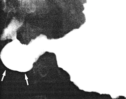
Case 34.1 G.C., l7 year old male, had a two year history of epigastric pain, vomiting,
weight loss and retardation of growth. Physical examination revealed severe iron
deficiency anaemia. Radiographic study showed multiple lobulated filling defects in the
corpus and sinus of the stomach, constant irregularity of the greater and lesser curvatures
and a narrowing at the commencement of the pyloric sphincteric cylinder, in the region of
the left pyloric loop (Fig. 34.1). The cylinder was partially contracted throughout the
examination, never contracting or relaxing maximally; this was associated with a patent
pyloric aperture measuring 4.0 mm in diameter. Gastric emptying of fluid barium was
delayed. Endoscopy showed diffuse, nodular infiltration of the entire corpus and
"antrum", with contact bleeding. The infiltration surrounded the pyloric orifice, which
was patent. The duodenum could not be visualized. Histology, according to the
"Working Formulation", revealed a high grade, malignant, non-Hodgkin lymphoma.
Bone marrow biopsy was normal.
 |
Fig. 34.1.
Case G.C. Irregularities of lesser and greater curvatures extending to
commencement of pyloric sphincteric cylinder. Cylinder partially contracted (arrows).
Pyloric aperture patent
|
At operation the entire stomach from the gastro-oesophageal junction to the pylorus was
found to be involved by lymphomatous infiltration, and a total gastrectomy with an
oesophago-jejunal anastomosis was performed. Macroscopically the resection specimen
showed diffuse thickening of the walls with effacement of the normal mucosal pattern.
Microscopically tumor cells extended from the mucosa into the muscular layer and in
several areas as far as the serosa. Electron microscopically the cells were determined to
belong to the lymphoma group, the condition being diagnosed by a combination of light
microscopy, electron microscopy and immunocytology as a large cell (B-cell),
immunoblastic lymphoma. The distal border of the resection specimen was free of tumor
cells and liver biopsy was normal. A few enlarged mesenteric lymph nodes proved to be
of reactive type.
Case 34.2 J.J., 43 year old male, presented with intermittent epigastric pain,
vomiting, loss of appetite and loss of weight, of one year's duration. Physical
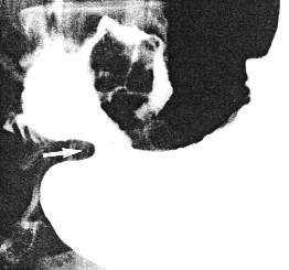
examination revealed epigastric tenderness. Radiographic study showed a constant
irregularity of the lower part of the lesser curvature, with a permanent, ulcer-like
projection; a large, lobulated filling defect was present in the duodenal bulb (Fig. 34.2).
The pyloric sphincteric cylinder remained expanded throughout the examination, failing
to contract, and the pyloric orifice remained patent. The appearances were ascribed to a
mass lesion, probably with ulceration, infiltrating the distal lesser curvature of the
stomach and the first part of the duodenum. At endoscopy a deformity of the gastric
"antrum", presumably due to external compression was seen, while the pyloric ring
appeared normal. A large polypoid, lobulated mass measuring approximately 4.0 cm in
diameter, was present in the duodenal bulb; the surrounding duodenal mucosa appeared
normal. The biopsy specimen was inadequate but the possibility of malignant lymphoma
was mentioned. Abdominal sonography showed no abnormality in the pancreas, liver,
gall bladder and kidneys. Chest radiographs and bone marrow trephine biopsies were
normal.
 |
Fig. 34.2
Case J.J. Irregularity lower lesser curvature with ulcer-like projection. Pyloric
sphincteric cylinder expanded. Pyloric aperture patent (arrow). Large, lobulated filling
defect in duodenal bulb
|
Previous Page | Table of Contents | Next Page
© Copyright PLiG 1998


