

Go to chapter: 1 | 2 | 3 | 4 | 5 | 6 | 7 | 8 | 9 | 10 | 11 | 12 | 13 | 14 | 15 | 16 | 17 | 18 | 19 | 20 | 21 | 22 | 23 | 24 | 25 | 26 | 27 | 28 | 29 | 30 | 31 | 32 | 33 | 34 | 35 | 36 | 37 | 38 | 39
Chapter 30 (page 149)
Holt et al. (l986) pointed out that gastric emptying rate measurements in patients with
duodenal ulcer had produced conflicting results. Many previous studies had failed to
identify any gastric emptying abnormality, while in others the rate was found to be faster
than in normal controls. The conflicting results might have been due to factors such as
differences in experimental methods and differences in meal composition; the exact
stage at which emptying was measured in relation to the treatment or healing of a
duodenal ulcer was also important. Using 113m In as a marker of the liquid,
and 99m Tc as a marker of the solid phase, measurements of gastric emptying
rates were undertaken in duodenal ulcer patients before, during and after therapy with
cimetidine, as well as in control subjects. Before therapy there were no significant
differences in the rate or pattern of gastric emptying in duodenal ulcer patients as
compared with normal controls. This implied that gastric motor function in the two
groups was similar, and the results did not agree with the findings of previous
investigators who had found abnormally fast emptying in duodenal ulcer patients.
During treatment, however, the emptying rate of particles became faster. The findings
suggested that cimetidine had no marked effect on the emptying of the liquid component
of a meal, but that there was a specific effect of the emptying of the solid phase. Gastric
emptying patterns from control subjects and from healed duodenal ulcer patients were
remarkably similar.
Williams et al. (l986) measured gastric emptying of citric acid, glucose and fat meals by
means of a double-sampling, dye-dilution technique, while maintaining intragastric pH at
a constant level. Acid, glucose and fat inhibited gastric emptying in a dose-dependent
fashion in duodenal ulcer patients as well as in normal controls. Duodenal ulcer patients
emptied all three types of meals faster than normals, but differences only occurred at the
lower doses of glucose or with the less potent doses of acid and fat. It was concluded that
differences in gastric emptying of liquid meals in duodenal ulcer patients, as compared
with normals, were small; the variable responses obtained with different concentrations
might explain some of the inconsistencies found by previous workers.
Hui et al. (l986) performed endoscopic biopsies in 213 patients with active duodenal
ulceration and diagnosed active chronic antral gastritis in 99 percent. The degree of
chronic inflammation was assessed histologically by the infiltration of polymorphs and
chronic inflammatory cells and by the severity of mucosal degeneration. In the majority
of patients antral gastritis was considered to be of a moderate degree. In non-ulcer
controls active chronic antral gastritis occurred in 50 percent and in a milder form. It was
concluded that the exact relationship of chronic antral gastritis to duodenal ulceration was
uncertain. Healing of the duodenal ulcer was accompanied by histological improvement
of the antral gastritis.
We examined the contractile behaviour of the pyloric sphincteric cylinder in duodenal
ulceration by means of upper gastrointestinal radiography in the following groups of
patients:
Group 1. Sixty consecutive patients with active duodenal ulceration receiving and
responding to medical treatment. All had been referred from the outpatient department
with the clinical diagnosis of duodenal ulcer. In most cases the symptoms had been
present for several months to a year; some had had symptoms and signs of chronic,
recurrent duodenal ulceration and had received anti-ulcer therapy intermittently for the
previous one to four years. Many had previous radiographic and/or endoscopic
examinations, in which the diagnosis had been confirmed. Only patients in whom a
definite ulcer niche could be seen radiologically or endoscopically in the duodenal bulb,
were admitted to the study; patients with additional upper gastrointestinal pathology, e.g.
gastric ulceration or hiatus hernia, were excluded.
Group 2. Seventeen cases of chronic recurring duodenal ulceration not responding
satisfactorily to medical therapy. All had endoscopically proven active duodenal
ulceration and all were candidates for the operation of sero-myotomy and parietal cell
vagotomy.
In 58 of 60 cases of the first group, and in 15 of l7 cases of the second group,
contractions of the pyloric sphincteric cylinder were normal. The following are examples
from the 58 cases of the first group:
Case Reports
Case 30.1. P.K., 44 year old male, a known case of duodenal ulceration, had received
anti-ulcer therapy for some months; at the time of examination he was still symptomatic.
Radiographic examination revealed no lesion in the oesophagus and stomach; the
duodenal bulb was deformed and showed a niche on its lesser curvature side, indicating
an active duodenal ulcer. Contractions of the pyloric sphincteric cylinder were normal.
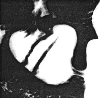
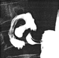
The distended cylinder contained several transverse (i.e. circular) mucosal folds (Fig.
30.1A). During contraction of the cylinder the folds changed in direction to
longitudinal, only longitudinal folds being evident in the maximally contracted cylinder,
with formation of the pyloric canal (Fig. 30.1B). The rate of cyclical
contractions of the cylinder was normal at about 3 or 4 per minute. It is not possible to
determine the amplitude of individual contractions radiographically; descriptions such as
"maximal" or "near maximal" may give some indication of the degree of contraction.
Radiographically the range and degree of contraction appeared to be normal.
A | B |
| Fig. 30.1 A,B.
Case P.K. A Transverse (i.e. circular) mucosal folds in distended pyloric sphincteric
cylinder. Ulcer niche in duodenal bulb. B Normal contraction of sphincteric cylinder.
Longitudinal mucosal folds in fully contracted pyloric canal. Ulcer niche in duodenal bulb
|
Previous Page | Table of Contents | Next Page
© Copyright PLiG 1998


