


Go to chapter: 1 | 2 | 3 | 4 | 5 | 6 | 7 | 8 | 9 | 10 | 11 | 12 | 13 | 14 | 15 | 16 | 17 | 18 | 19 | 20 | 21 | 22 | 23 | 24 | 25 | 26 | 27 | 28 | 29 | 30 | 31 | 32 | 33 | 34 | 35 | 36 | 37 | 38 | 39
Chapter 29 (page 145)
Lawson (l98l) stated that a prepyloric ulcer differed from a duodenal or mid lesser
curvature gastric ulcer in a number of ways. For instance, it had a high recurrence rate
when treated by parietal cell vagotomy and it was associated with a variable level of
gastric acid secretion. The degree and distribution of chronic severe gastritis in 15
antrectomy specimens removed for prepyloric ulceration was examined. In 6 of the cases
the prepyloric ulcer was on the lesser curvature; in these cases there were localized areas
of severe chronic gastritis along the middle of the anterior and posterior walls of the
"antrum", with a tongue of gastritis extending upwards along the lesser curvature as far as
the fundus. In 3 cases, where the prepyloric ulcer was situated on the greater curvature,
localized areas of gastritis also occurred, the least affected area being along the lesser
curvature. The widest area of gastritis was seen on the anterior and posterior walls of the
greater curvature of the "antrum", with a tongue-like extension to the fundus. In 6 cases
the ulcer had healed at the time of histological examination. The general pattern of the
distribution of severe gastritis was similar to that in the 9 other cases, the main area
affected being the lesser curvature in both the fundus and antrum. It was concluded that
localized areas of severe chronic gastritis occurred in the "antrum" in all cases of
prepyloric ulceration. A tongue-like process of gastritis extended from the "antrum" to
the fundus along the particular curvature on which the ulcer was situated. In prepyloric
ulceration not the whole of the "antrum" was involved, as appeared to be the case in
many specimens resected for mid lesser curvature gastric ulcer. The fact that the mucosa
failed to return to normal after healing of the ulcer in 6 cases, confirmed the previous
findings of Gear et al. (l97l), and suggested that chronic gastritis was not a secondary
effect of the ulcer but could be an important factor in its pathogenesis.
Brooks (l985) found that patients with pyloric and duodenal ulcers showed a similar
tendency to hypersecretion, but that peak acid outputs were significantly lower in the
prepyloric ulcer group.
Lauritsen (l988, l989) stated that although it was generally believed that prepyloric ulcers
resembled duodenal ulcers with respect to their acid secretory patterns, clinical trials had
shown that patients with prepyloric ulcer disease were more resistant to treatment with
H2-receptor antagonists than patients with duodenal ulcer disease. Their treatment
remained a therapeutic challenge; for that reason the effects of a proton pump inhibitor
(omeprazole) were studied in 176 patients with active prepyloric ulcers (all gastric ulcers
below the angulus were considered to be "prepyloric"). The results were said to be
encouraging.
The following are representative cases of ulceration within the pyloric sphincteric
cylinder:
Case Reports
Case 29.5 S.S., 32 year old female, complained of heartburn and intermittent
epigastric pain of two years' duration. Radiographic examination showed a constant fleck
of barium, indicating an active ulcer, within the pyloric sphincteric cylinder
approximately 1.5 cm proximal to the pyloric aperture (Fig. 29.5). The cylinder was
partially contracted, and remained contracted throughout the examination, with absence
of normal, cyclical contraction and relaxation. Emptying of fluid barium was not
delayed. Anti-ulcer therapy was commenced and proved beneficial. Endoscopy 5
months later showed a superficial, healing gastric ulcer in the anterior wall of the pyloric
region, without evidence of malignancy. Follow-up endoscopy showed healing of the
ulcer. The duodenum was normal. Considerable duodenogastric reflux was present.
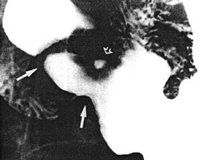 |
Fig. 29.5.
Case S.S. Ulcer (open arrow) within confines of pyloric sphincteric cylinder, which is
partially contracted (filled arrows)
|
Case 29.6 M.T., 20 year old male, complained of dyspepsia, loss of appetite and
epigastric pain of several months' duration. Radiographic examination showed a constant
fleck of barium, 1.5 cm in diameter, indicating an active ulcer, on the lesser curvature
side of the pyloric sphincteric cylinder (Fig. 29.6). The cylinder was somewhat
deformed, partially contracted and unchanging in appearance throughout the
examination, with absent cyclical activity; it contained prominent oblique and circular
mucosal folds which were drawn in towards the ulcer and did not change in direction
(Fig. 29.6). The duodenal bulb filled poorly. Response to anti-ulcer therapy was poor.
Endoscopy 3 months later confirmed the presence of a benign gastric ulcer in the pyloric
region. Billroth I partial gastrectomy with truncal vagotomy in May l984 showed a
chronic, benign gastric ulcer 1.0 cm proximal to the pylorus. Microscopic examination
revealed absence of malignancy. The gastric mucosa showed evidence of chronic
gastritis.
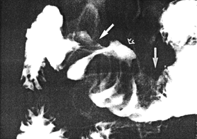 |
Fig. 29.6.
Case M.T. Ulcer (open arrow) in sphincteric cylinder, which is deformed and
permanently contracted (filled arrows). Circular, unchanging mucosal folds drawn in
towards ulcer
|
Previous Page | Table of Contents | Next Page
© Copyright PLiG 1998