


Go to chapter: 1 | 2 | 3 | 4 | 5 | 6 | 7 | 8 | 9 | 10 | 11 | 12 | 13 | 14 | 15 | 16 | 17 | 18 | 19 | 20 | 21 | 22 | 23 | 24 | 25 | 26 | 27 | 28 | 29 | 30 | 31 | 32 | 33 | 34 | 35 | 36 | 37 | 38 | 39
Chapter 29 (page 142)

Go to chapter: 1 | 2 | 3 | 4 | 5 | 6 | 7 | 8 | 9 | 10 | 11 | 12 | 13 | 14 | 15 | 16 | 17 | 18 | 19 | 20 | 21 | 22 | 23 | 24 | 25 | 26 | 27 | 28 | 29 | 30 | 31 | 32 | 33 | 34 | 35 | 36 | 37 | 38 | 39
Chapter 29 (page 142)
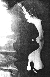 |  |
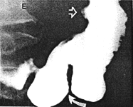
| Fig. 29.2 A,B. A Case I.H. Large gastric ulcer posterior wall of corpus (open arrow). Deep spastic incisura distal greater curvature (curved arrow). B Case I.H. The spastic incisura is caused by constant contraction of the left pyloric loop (curved arrow). | |
 |  |
 | 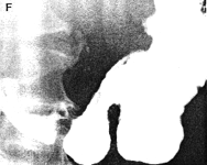 |
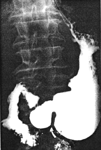
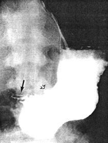
| Fig. 29.2 C-F Case I.H. Eighteen months later the gastric ulcer (open arrow), contraction of the left pyloric loop (curved arrow) and contraction of the sphincteric cylinder are unchanged | |
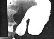
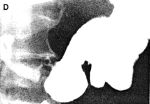
Case 29.3 V.D., 30 year old male, complained of epigastric pain not responding to antacids. Radiographic examination showed a gastric ulcer 1.5cm in diameter, on the lesser curvature at the angulus (Fig. 29.3A); no signs of malignancy were seen. Intially there was marked contraction of the pyloric sphincteric cylinder, which contained a few prominent mucosal folds; this was associated with delay in emptying of liquid barium. After barium had filled the cylinder, it was seen to remain contracted throughout the examination; although a minor degree of relaxation occurred occasionally, the apperances remained as illustrated most of the time, with absence of cyclical activity (Fig. 29.3B). Longitudinal mucosal folds were evident in the contracted cylinder. Endoscopic biopsy showed a benign looking gastric ulcer in the corpus. Microscopically there was mixed inflammatory cell infiltration, diagnosed as acute on chronic gastritis. No evidence of malignancy was seen. Repeat endoscopy 2 months later showed that the ulcer had healed.
 A A | |
| Fig. 29.3. A Case V.D. Gastric ulcer on lesser curvature at angulus (open arrow). Marked contraction of sphincteric cylinder (filled arrow). B Case V.D. Gastric ulcer at angulus (open arrow). Constant contraction of sphincteric cylinder (filled arrows) with absent cyclical activity. | 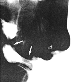 B B |
Previous Page | Table of Contents | Next Page
© Copyright PLiG 1998