

Go to chapter: 1 | 2 | 3 | 4 | 5 | 6 | 7 | 8 | 9 | 10 | 11 | 12 | 13 | 14 | 15 | 16 | 17 | 18 | 19 | 20 | 21 | 22 | 23 | 24 | 25 | 26 | 27 | 28 | 29 | 30 | 31 | 32 | 33 | 34 | 35 | 36 | 37 | 38 | 39
Chapter 27 (page 126)
Ehrlein (l98l) pointed out that the function of the pyloric sphincter in preventing
duodenogastric reflux had not been clarified. (It was explained that the term "pyloric
sphincter" in this context referred to the pyloric ring). It was difficult to record
contractions over a long period of time in humans. In animals it was possible to measure
the external diameter of the pylorus with induction coils, and to determine pressure
changes simultaneously by means of implanted strain gauge transducers (Ehrlein l980).
The method was used in canines to investigate possible co-ordination of gastric and
duodenal motility, gastroduodenal reflux at the same time being observed
fluoroscopically. In the digestive state the pylorus opened when a peristaltic wave
involved the commencement of the "antrum", closing when the wave reached the pyloric
sphincter. Contraction maxima of the duodenal bulb most often occurred a fraction of a
second before, or after, contraction maxima of the pyloric sphincter. Consequently there
was incomplete closure of the pylorus during duodenal contraction, allowing
duodenogastric reflux to occur; it was produced by atypical segmental contractions of
the bulb. In the interdigestive state different phases were encountered. In the first phase,
in the absence of contractile activity in the stomach and duodenum, no reflux occurred.
In the second and third phases imperfect timing of pyloric and duodenal contractions, as
in the digestive state, sometimes resulted in duodenogastric reflux.
Various derivatives of 99mTc-IDA have now been used extensively in the study
of duodenogastric reflux, notably 99mTc-BIDA (butyl iminodiacetic acid) and
99mTc-HIDA. These tests have certain minor limitations in common (Thomas et
al, l984). For instance, anatomical definition of the stomach was often complicated by
overlap of the left lobe of the liver and the duodeno-jejunal flexure; delineation of the
gastric "antrum" was poor, and as it was close to the duodenal bulb, minor degrees of
reflux might not be detectable; and computer analysis of small volumes of reflux might
be difficult to interpret.
Two further limitations of the cholescintigraphic tests, in our view, are the following:
- As only bile constituents are labelled, the tests apply to reflux of bile into the
stomach; they are not a measure of reflux of the other constituents of duodenal
juice, of which pancreatic exocrine secretion is the most important. It may be
useful to have a test able to demonstrate reflux of duodenal contents (as opposed
to bile only), into the stomach.
- At present the tests do not visualize the pylorus clearly, i.e. it is not possible to
determine the extent of contraction or relaxation of the pyloric region in relation
to reflux.
As a modification of the double-contrast upper gastrointestinal radiographic examination,
we have described the following radiographic test for duodenogastric reflux (Keet l982).
In this test the pylorus is clearly visualized and its diameter can be determined during
reflux; in addition, the relationship of reflux to pyloric and duodenal contractions may be
studied. The test is non-invasive and does not entail the use of catheters, intubation or the
administration of pharmacological agents (which may alter motility).
The patient, standing behind the radiological TV monitor after a 12-hour overnight fast,
is instructed to swallow 4 to 5 mouthfuls of a micropulverized barium suspension, e.g.
Micropaque (Adcock-Ingram, Johannesburg) ordinarily used for upper gastrointestinal
radiographic examinations. Immediately afterwards a gas-producing agent is swallowed,
e.g. 2 x 50 Gastrast tablets (Toho Kagaku Kenyusho, Tokyo), followed by 2 mouthfuls of
water containing a few drops of Telament liquid (Adcock Ingram, Johannesburg). The
barium accumulates in the lower part of the stomach while the gas distends the fornix.
While the patient is instructed not to eructate, the table is immediately tilted into the
horizontal position. With the arms abducted throughout the examination, the supine
subject is now rotated into the left anterior oblique position (right side down) till barium
enters the duodenal bulb. As soon as duodenal filling is achieved, the subject is rotated
rapidly through 90 degrees into the right anterior oblique position. This causes the
remaining barium in the stomach to descend into the fornix, while the gas is displaced
and ascends into the pyloric region, which now constitutes the uppermost part of the
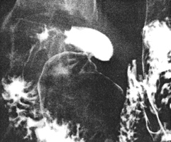
stomach. Consequently the first part of the duodenum is filled with barium, while the
pyloric region up to the ring is filled with gas (Fig. 27.1). The competence of the pylorus
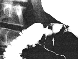
can now be studied, radiographs being taken at appropriate times. Duodenogastric reflux
through the pylorus into the gas-containing part of the gastric lumen is clearly visible
(Fig. 27.2). (This should not be confused with the normal orad movement of barium
often seen during contraction of the pyloric sphincteric cylinder as described in Chap.
13). Should no reflux be observed, or should the duodenum empty its contents
prematurely, positioning of the patient is repeated. Generally speaking, the manoeuvre is
repeated 4 to 5 times during each test. The effects of compression of the second or third
parts of the duodenum may also be studied.
 |
Fig. 27.1.
Duodenal bulb filled with barium. Pyloric sphincteric cylinder filled with gas
and distended
|
 |
Fig. 27.2.
Reflux of barium (arrow) from the duodenum into the pyloric sphincteric
cylinder, which is relaxed
|
Previous Page | Table of Contents | Next Page
© Copyright PLiG 1998


