


Go to chapter: 1 | 2 | 3 | 4 | 5 | 6 | 7 | 8 | 9 | 10 | 11 | 12 | 13 | 14 | 15 | 16 | 17 | 18 | 19 | 20 | 21 | 22 | 23 | 24 | 25 | 26 | 27 | 28 | 29 | 30 | 31 | 32 | 33 | 34 | 35 | 36 | 37 | 38 | 39
Chapter 22 (page 101)


Go to chapter: 1 | 2 | 3 | 4 | 5 | 6 | 7 | 8 | 9 | 10 | 11 | 12 | 13 | 14 | 15 | 16 | 17 | 18 | 19 | 20 | 21 | 22 | 23 | 24 | 25 | 26 | 27 | 28 | 29 | 30 | 31 | 32 | 33 | 34 | 35 | 36 | 37 | 38 | 39
Chapter 22 (page 101)
A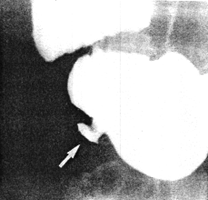 | B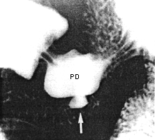 |
| Fig. 22.1. A Case M.W. Small intramural diverticulum (arrow) on greater curvature of pyloric sphincteric cylinder 2.5 cm proximal to pyloric ring. B Case M.W. Partial contraction of sphincteric cylinder. Intramural diverticulum (arrow) situated on rim of physiological pseudodiverticulum (PD), midway between right and left pyloric loops | |
Because of the features enumerated in the discussion it was diagnosed as an intramural or
partial gastric diverticulum. It was certainly not an ulcer as witnessed by repeated
gastroscopies. The fact that it was not seen at gastroscopy does not come as a surprise as
Treichel et al. (l976) had found that the lesion was easier to detect by radiography than by
endoscopy. Cockrell et al. (l984) stated that an intramural diverticulum may be difficult
to detect endoscopically if its ostium is hidden by a fold or if it occurs in an area which is
contracting.
Case 22.3. J.V., l9 year old female, complained of "acidity", of several months'
duration. Gastroscopy showed mucosal erosions in the lower oesophagus, probably due
to reflux oesophagitis. The gastric fornix and corpus were normal; on the greater
curvature of the "antrum" a "pseudodiverticulum" was seen. Radiographic examination
14 months later, for the same complaint, showed an intramural diverticulum on the
greater curvature within 2.0 cm of the pylorus. Cyclical contractions of the pyloric
sphincteric cylinder were normal; during contraction of the right and left pyloric loops, a
normal, physiological pseudodiverticulum was seen with an intramural diverticulum on
its greater curvature aspect (Fig. 22.3).
Previous Page | Table of Contents | Next Page
Case 22.2. G.V., 32 year old female, complained of a vague feeling of fullness and
occasional pain in the epigastrium. Physical examination revealed some epigastric
tenderness. After a month's treatment with antacids the symptoms disappeared.
Radiographic examination at that time showed a small diverticular-like structure on the
greater curvature of the pyloric sphincteric cylinder approximately 3.0 cm proximal to the
pyloric ring, i.e. midway between the right and left pyloric loops, in the position where
the pyloric pseudo-diverticulum usually occurs (Fig. 22.2
A 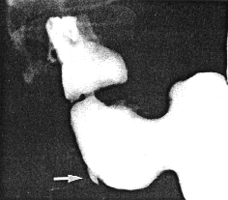
B 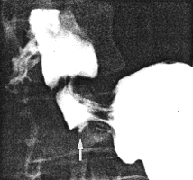
Fig. 22.2. A
Case G.V. Intramural diverticulum (arrow) on greater curvature
of sphincteric cylinder 3.0 cm proximal to pyloric ring.
B Case G.V. Contraction of sphincteric cylinder. Intramural
diverticulum (arrow) between right and left pyloric loops now smaller
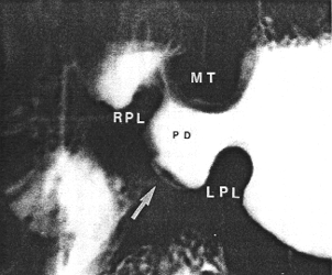
Fig. 22.3.
Case J.V. Normal contraction of pyloric muscle torus (MT) and right (RPL)
and left (LPL) pyloric loops. Intramural diverticulum (arrow) on rim of physiological
pseudodiverticulum (PD)
© Copyright PLiG 1998