


Go to chapter: 1 | 2 | 3 | 4 | 5 | 6 | 7 | 8 | 9 | 10 | 11 | 12 | 13 | 14 | 15 | 16 | 17 | 18 | 19 | 20 | 21 | 22 | 23 | 24 | 25 | 26 | 27 | 28 | 29 | 30 | 31 | 32 | 33 | 34 | 35 | 36 | 37 | 38 | 39
Chapter 21 (page 99)
Samuel (l955) pointed out that barium-filled ducts in ectopic pancreatic tissue, embedded
in the wall of the stomach, may simulate an intramural diverticulum. Moore and Kaplan
(l956) found that a small mass of aberrant pancreatic tissue may be loosely attached to
the serosal surface of the upper intestine, or it may occur in the subserosal, submuscular
or submucosal regions of the gastric walls. Radiographically it usually presents as an
elevated mound with a central dimpling and an opening formed by ducts. When the
dimpling is filled with barium, it may resemble a polyp with an ulcer at its tip; when the
ducts are filled, it resembles a diverticulum.
Besemann et al. (l97l) confirmed that most aberrant pancreatic nodules occurred in the
upper gastrointestinal tract, usually in the duodenum, but occasionally in the stomach
near the pylorus.
Case 21.1. D.J., 22 year old male, had a long history of dyspepsia not responding to
anti-ulcer treatment. Two days before admission there had been an episode of
haematemesis. Gastroscopy showed a small orifice on the posterior gastric wall close to
the pyloric aperture. It was thought to be either an intramural diverticulum or a double
pylorus; radiographic examination was requested for further elucidation. This showed a
small, branching, diverticular structure approximately 1.0 cm in length, on the greater
curvature side of the pyloric sphincteric cylinder within 1.0 cm of the pylorus (Fig. 21.1).
It remained constant in appearance irrespective of the degree of contraction of the
cylinder, the contractions being normal. At laparotomy no evidence of ulceration was
seen. A nodule of abnormal tissue measuring approximately 2.0 cm in diameter was
palpated in the gastric wall at the site indicated by endoscopy and radiology.
Gastrostomy revealed a small orifice extending into the nodule, which was removed by
local excision. Histology showed it to consist of endo- and exocrine pancreatic tissue
located in the muscular coat of the stomach.
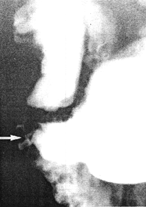 |
Fig. 21.1.
Case D.J. Small, branching, diverticulum-like structure (arrow) on greater
curvature side of pyloric sphincteric cylinder
|
In this case a small nodule of ectopic pancreatic tissue in the wall of the greater curvature
of the pyloric sphincteric cylinder did not affect the movements of the cylinder to any
appreciable extent. Radiographically the lesion mimicked an intramural diverticulum
(Chap. 22).
- Besemann EF, Auerbach SH, Wolfe WW. Importance of roentgenologic
diagnosis of aberrant pancreatic tissue in the gastrointestinal tract. Amer J
Roentg Rad Ther Nucl Med l969, 107, 71-76.
- Moore MB, Kaplan IW. Heterotopic pancreatic tissue in the stomach.
Amer J Gastroenterol l956, 26, 699-705.
- Samuel E. Gastric diverticula. Brit J Radiol l955, 28, 574-578.
Previous Page | Table of Contents | Next Page
© Copyright PLiG 1998