


Go to chapter: 1 | 2 | 3 | 4 | 5 | 6 | 7 | 8 | 9 | 10 | 11 | 12 | 13 | 14 | 15 | 16 | 17 | 18 | 19 | 20 | 21 | 22 | 23 | 24 | 25 | 26 | 27 | 28 | 29 | 30 | 31 | 32 | 33 | 34 | 35 | 36 | 37 | 38 | 39
Chapter 20 (page 92)
Direct viewing of the pyloric musculature is achieved by means of ultrasonic examination
(Chap. 10). Blumhagen and Coombs (l98l) showed that a persistent contraction of the
circular musculature of the "distal antrum and pylorus", creating a cylindrical muscle
mass resembling IHPS, might be seen during ultrasonic examinations in infants.
According to these authors, contraction of the "distal antrum" and pylorus (and not of the
pyloric ring) constituted pylorospasm.
Blumhagen and Noble (l983) found that spasm of the "distal antrum and pylorus" could
be differentiated ultrasonically from IHPS by measuring the single wall muscle thickness.
Normally the muscle thickness in infants varies between 1.5 and 3.0 mm; in IHPS it
varies between 3.0 and 6.0 mm. Whenever the single wall muscle thickness is 4.0 mm or
more, it can be regarded as IHPS.
Wilson and Vanhoutte (l984) determined the true length of the pyloric muscle by means
of ultrasonography in l7 normal infants; the range varied from 12.0 to 15.0 mm. Similar
results were obtained by Graif et al. (l984), who found the mean length of the pylorus
(pyloric musculature) to be 12.0 mm with a standard deviation of 3.7 mm. It is clear that
this is the anatomical region which may contract during pylorospasm, i.e. a relatively
long area which includes the pyloric ring, as opposed to an isolated contraction of the
ring only. This was confirmed by Stunden et al. (l986), who measured the length of the
pyloric canal in normal infants "with the pylorus in its most contracted state". The mean
length of the canal was 8.3 ± 2.5mm, and on occasion it was up to 14.0 mm in
length.
With longitudinal ultrasonic views of the pylorus Haller and Cohen (l986) found the
length of the pyloric musculature in normal infants to be up to l8.0 mm. Ultrasonically
the contraction of this muscular cylinder in pylorospasm could simulate IHPS. However,
with spasm the contracted region was not quite as rigid as in IHPS, and the appearance
changed on subsequent examinations.
Larson et al (l967) described 10 adult cases in whom radiographs showed clear evidence
of adult hypertrophic pyloric stenosis (AHPS), i.e. a cylindrical pyloric narrowing (Chap.
24). At operation the stomach was found to be normal in 5, showing that the narrowing
was of a temporary, spastic nature, in other words due to pylorospasm.
During operations on 5 suspected cases of AHPS, Bateson et al. (l969) noted that on
touching the pylorus with a gloved finger, the pyloric and adjacent "antral" muscle
contracted, becoming hard and pale; the affected region was approximately 2.5 cm in
length and resembled AHPS, but the contraction was temporary and of a spastic nature.
Keet and Heydenrych (l97l) found that electrical and mechanical stimulation of the vagus
trunks in the oesophageal hiatus of the diaphragm in canines, caused a 3.0 cm long
cylindrical area of contraction in the pyloric region. It corresponded exactly to the
anatomical pyloric sphincteric cylinder as described by Cunningham (l906), Forssell
(l913) and Torgersen (l942), lasted as long as the stimulus was applied, and was clearly
of a spastic nature.
The following are some of the cases of pylorospasm which we have encountered:
Case 20.2. R.G., 10 day old male infant was admitted for persistent vomiting. Upper
gastrointestinal barium study showed a constant, string-like narrowing 2.0 cm in length in
the pyloric region, clearly limited to the sphincteric cylinder (Fig. 20.2). It was
associated with a concave indentation of the base of the duodenal bulb, the appearance
resembling infantile hypertrophic pyloric stenosis. At laparotomy no pyloric tumor was
found. There was some kinking of the duodenum, which was corrected, and in view of
the radiologic findings a pyloromyotomy was done. A naso-jejunal tube was inserted and
recovery was uneventful. The final diagnosis of the pyloric narrowing was spasm of the
pyloric sphincteric cylinder.
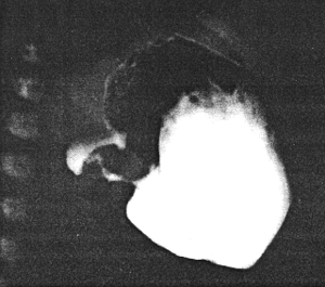 |
Fig. 20.2.
Case R.G. Constant spasm of pyloric sphincteric cylinder, simulating infantile
hypertrophic pyloric stenosis. At operation no muscular hypertrophy was found
|
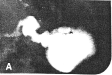
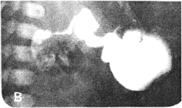
Case 20.3. B.F., 7 day old female infant presented with persistent, bile-stained
vomiting since birth. Radiological study showed a tubular narrowing 2.0 cm in length in
the pyloric region (Fig. 20.3). It conformed to partial contraction of the pyloric
sphincteric cylinder with a prominent impression of the pyloric muscle knot on the lesser
curvature side. There was total absence of cyclical contraction and relaxation of the
cylinder, the pyloric aperture being fixed in the open or patent position. The duodenal
cap was normal but there appeared to be some narrowing of the remainder of the first part
of the duodenum. Infantile hypertrophic pyloric stenosis could not be excluded.
Laparotomy revealed partial obstruction at the duodeno-jejunal junction due to ectopic
pancreatic tissue in the intestinal wall and adhesions. The pylorus showed no organic
lesion. After biopsy, severance of adhesions and pyloromyotomy, recovery was
complete. The pyloric narrowing was diagnosed as spasm of the sphincteric cylinder.
 |  |
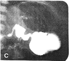 | 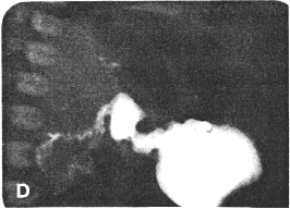 |
| Fig. 20.3 A-D.
Case B.F. Constant contraction of sphincteric cylinder. At operation no pyloric
lesion was found
|
Previous Page | Table of Contents | Next Page
© Copyright PLiG 1998