


Go to chapter: 1 | 2 | 3 | 4 | 5 | 6 | 7 | 8 | 9 | 10 | 11 | 12 | 13 | 14 | 15 | 16 | 17 | 18 | 19 | 20 | 21 | 22 | 23 | 24 | 25 | 26 | 27 | 28 | 29 | 30 | 31 | 32 | 33 | 34 | 35 | 36 | 37 | 38 | 39
Chapter 11 (page 45)
Microscopic Anatomy
Illustrations of microscopic sections of the normal pyloric ring are occasionally
encountered in published papers. In most instances the purpose is to illustrate some
feature of the stomach in the vicinity of the pylorus and the ring is seen incidentally.
On examining such illustrations the anatomical build of the ring may be studied.
Horton (l928) illustrated 3 normal stomachs sectioned 6 hours post-mortem. In all it
is seen that the pyloric ring is not a structure consisting solely of muscular tissue.
While its "base" is formed by muscularis externa, the top or inner part of the ring( i.e.
the part surrounding the aperture) consists of a mucosal fold (made up of a core of
submucosa with a layer of mucosa on each surface, as described in Chapter 5). In
Horton's illustration of a 6 months old foetus the height of the outer muscular
component of the pyloric ring is 14.0mm and the height of the inner mucosal fold, or
mucosal component, also 14.0mm. In a 4 month old infant the muscular division of
the ring measures 3.4cm in height and the mucosal division 2.0cm. In a one year old
child the measurements are 3.5cm and 1.5cm respectively. It is clear in these subjects
that the inner part of the pyloric ring consists of mucosal and submucosal, and not of
muscular tissue.
Cole (l928) held that the abrupt interruption separating the lumen of the stomach from
that of the duodenum (i.e. the pyloric ring) is a muco-membranous fold consisting
solely of mucosa and submucosa with its acompanying muscularis mucosae. The
circular fibres of the muscularis externa end at the base of the "pyloric fold", which
should be looked upon as a valve and not as a sphincter, according to Cole (l928).
Scott's (l946) observations led him to conclude that normally a thick mucosal fold
caps the pyloric "sphincter" (from which it may be concluded that the ring consists of
both the "sphincter" and an overlying mucosal fold).
In illustrations of Manning and Gunter (l950) the following measurements may be
made: in a 66 year old subject the external muscular component of the ring is 3.0cm
in height, and the inner mucosal/submucosal component 2.5 cm. In a 78 year old
subject the figures are 2.5cm and 2.0cm respectively, and in a 58 year old subject
3.5cm and 2.0cm respectively. Williams (l962) illustrated two fresh, adult, partial
gastrectomy specimens. In one the muscular component of the ring measures l8.0mm
in height and the mucosal/submucosal component 9.0mm. In the other the figures are
21.0mm and 11.0mm respectively.
Although the nature and function of the pyloric ring is often debated, few deliberate
attempts have been made to determine its microscopic anatomy. Consequently
sections of the ring were done more than 6 hours post-mortem in 7 subjects varying in
age from 9 months to 83 years, who had succumbed to non-gastrointestinal causes; a
total of 20 sections were examined (Table 11.2). In all cases the pyloric ring is seen
to consist of muscular and mucosal/submucosal components (Fig 11.3). For instance,
in a 9 month old subject the muscular component is seen to be 2.0mm in height, and
the overlying mucosal component 1.0mm. In a 5 year old subject the approximate
figures are 5.5mm and 1.5mm respectively, and in a 44 year old subject 5.5mm and
1.5mm respectively.
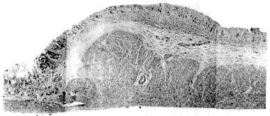 |
| Fig. 11.3. Microscopic section of
normal pyloric ring. The muscular component measures 4.5 mm, the mucosal/submucosal component 1.5 mm
in height |
Table 11.2 Results of postmortem examination in seven subjects
|
| Subject | Age | Sex | Specimen number | Total wall thickness (mm) |
Thickness of muscle layers | Thickness of mucosal/submucosal component |
|
| A.R. | 9m | M | 1 | 3.0 | 2.0 | 1.0 |
| | | 2 | 3.0 | 2.0 | 1.0 |
| | | 3 | 3.5 | 2.5 | 1.0 |
| C.J. | 5y | M | 4 | 7.0 | 5.5 | 1.5 |
| | | 5 | 6.0 | 5.0 | 1.0 |
| | | 6 | 6.5 | 5.5 | 1.0 |
| | | 7 | 7.0 | 5.5 | 1.5 |
| A.W. | 34y | M | 8 | 6.5 | 5.0 | 1.5 |
| | | 9 | 6.0 | 5.0 | 1.0 |
| D.J. | 44y | M | 10 | 7.0 | 5.5 | 1.5 |
| | | 11 | 7.0 | 5.5 | 1.5 |
| | | 12 | 7.0 | 5.5 | 1.5 |
| | | 13 | 6.0 | 5.0 | 1.0 |
| | | 14 | 6.5 | 5.0 | 1.5 |
| K.P. | 55y | F | 15 | 5.0 | 3.5 | 1.5 |
| | | 16 | 5.5 | 4.0 | 1.5 |
| | | 17 | 5.0 | 4.0 | 1.0 |
| L.S. | 76y | M | 18 | 11.0 | 9.0 | 2.0 |
| A.W. | 83y | F | 19 | 6.5 | 5.5 | 1.0 |
| | | 20 | 6.5 | 5.5 | 1.0 |
|
y = years
m = months
It is concluded that the pyloric ring consists of both muscular and
mucosal/submucosal components. In anatomical specimens the muscular component
accounts for approximately two-thirds of the total height of the ring, and the overlying
mucosal/submucosal component for one-third. It is known that post-mortem autolysis
of the mucosa sets in rapidly; Williams (l962) also pointed out that fixation
diminishes the turgescence of the mucosal folds, contracts the stomach and reduces
the submucosal space, so that sections after fixation are a poor representation of the
living state. It may be concluded that the mucosal/submucosal component of the ring
forms a more prominent part during life than would appear from microscopic
sections.
Previous Page | Table of Contents | Next Page
© Copyright PLiG 1998